International CME on Renal Pathology in India

This presentation includes two last viewed slides from the International CME on Renal Pathology held in India, covering topics such as renovascular and tubulo interstitial diseases. The coordinators and presenters are prominent professors and experts in the field of pathology.
- Uploaded on | 2 Views
-
 nashwa
About International CME on Renal Pathology in India
PowerPoint presentation about 'International CME on Renal Pathology in India'. This presentation describes the topic on This presentation includes two last viewed slides from the International CME on Renal Pathology held in India, covering topics such as renovascular and tubulo interstitial diseases. The coordinators and presenters are prominent professors and experts in the field of pathology.. The key topics included in this slideshow are Renal pathology, CME, India, renovascular diseases, tubulo-interstitial diseases,. Download this presentation absolutely free.
Presentation Transcript
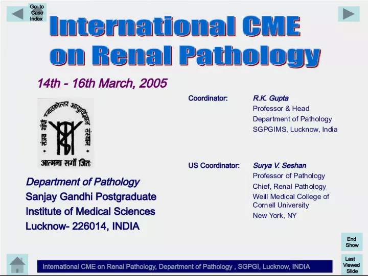
1. Last Viewed Slide International CME on Renal Pathology, Department of Pathology , SGPGI, Lucknow, INDIA End Show Go to Case Index 14th - 16th March, 2005 Coordinator: R.K. Gupta Professor & Head Department of Pathology SGPGIMS, Lucknow, India Department of Pathology Sanjay Gandhi Postgraduate Institute of Medical Sciences Lucknow- 226014, INDIA US Coordinator: Surya V. Seshan Professor of Pathology Chief, Renal Pathology Weill Medical College of Cornell University New York, NY
2. Last Viewed Slide International CME on Renal Pathology, Department of Pathology , SGPGI, Lucknow, INDIA End Show Go to Case Index Slide Seminar III Pathology of Renovascular & Tubulo-interstitial Diseases Sharda Sabnis Chief, Division of Renal Pathology Armed Forces Institute of Pathology Washington DC Lorraine Racusen Prof. of Pathology Johns Hopkins Medical School Baltimore, MD
3. Last Viewed Slide International CME on Renal Pathology, Department of Pathology , SGPGI, Lucknow, INDIA End Show Go to Case Index Case 1: 33 years gravida III with hypertension and generalized edema Case 2: 71 yrs female with hypertension, renal crisis, tight skin Case 3: 52 yrs male with flank pain, red urine, proteinuria Case 4: 52 yrs male with chronic eye problems and NSAID intake Case 5: 60 yrs female with atrial fibrillations and red urine Case 6: 33 yrs old female on anti-retroviral therapy Slide Seminar- III Pathology of Renovascular & Tubulo-interstitial Diseases
4. Last Viewed Slide International CME on Renal Pathology, Department of Pathology , SGPGI, Lucknow, INDIA End Show Go to Case Index Case 1 H/O 33 yrs gravida III, abortion I and para 1 presented at 24 weeks of pregnancy with elevated BP 160-170/100 mm Hg. Was normotensive during previous pregnancy. At 7 weeks patient had baseline hypertension with urinary protein of 111mg/day and normal LFTs. After 20 week gestation she noticed headache, followed by nausea, vomiting and neck discomfort. She had pleuritic chest pain, sore throat and diarrhea. A week later she noted significant generalized edema. She had significant proteinuria and positive ANA test. No H/O visual disturbances, current headaches, nausea, vomiting, abdominal pain, vaginal bleeding, joint pains, rashes, oral ulcerations. Slide Seminar- III: Pathology of Renovascular & TI Diseases - Case 1
5. Last Viewed Slide International CME on Renal Pathology, Department of Pathology , SGPGI, Lucknow, INDIA End Show Go to Case Index Case 1 P/E: BP 182/118 mm Hg, generalized edema, shortness of breath, genital herpes, abdominal exam normal for gestation, no tenderness or other findings. Laboratory findings: CBC: Hb 11.7, HCT 34, platelets 212000, white cell count 12.2 U/A: >300mg/dl protein (> 9gms/day), 10-25 RBCs/hpf with casts, 5-10 WBCs/hpf, urine output 30 cc/ hour BUN 19, Creatinine 0.9, bicarb 21, Glucose 118, albumin 2.3, total protein 5.1, ALT 51, AST41, Alk Phos 155, uric acid 7.6, anti-DNA antibody negative, complement levels-pending Slide Seminar- III: Pathology of Renovascular & TI Diseases - Case 1
6. Last Viewed Slide International CME on Renal Pathology, Department of Pathology , SGPGI, Lucknow, INDIA End Show Go to Case Index Slide Seminar- III: Pathology of Renovascular & TI Diseases - Case 1
7. Last Viewed Slide International CME on Renal Pathology, Department of Pathology , SGPGI, Lucknow, INDIA End Show Go to Case Index Slide Seminar- III: Pathology of Renovascular & TI Diseases - Case 1
8. Last Viewed Slide International CME on Renal Pathology, Department of Pathology , SGPGI, Lucknow, INDIA End Show Go to Case Index Slide Seminar- III: Pathology of Renovascular & TI Diseases - Case 1
9. Last Viewed Slide International CME on Renal Pathology, Department of Pathology , SGPGI, Lucknow, INDIA End Show Go to Case Index Slide Seminar- III: Pathology of Renovascular & TI Diseases - Case 1
10. Last Viewed Slide International CME on Renal Pathology, Department of Pathology , SGPGI, Lucknow, INDIA End Show Go to Case Index Slide Seminar- III: Pathology of Renovascular & TI Diseases - Case 1
11. Last Viewed Slide International CME on Renal Pathology, Department of Pathology , SGPGI, Lucknow, INDIA End Show Go to Case Index Slide Seminar- III: Pathology of Renovascular & TI Diseases - Case 1
12. Last Viewed Slide International CME on Renal Pathology, Department of Pathology , SGPGI, Lucknow, INDIA End Show Go to Case Index Case 1 ? Diagnosis Slide Seminar- III: Pathology of Renovascular & TI Diseases - Case 1
13. D I A G N O S I S & D I S C U S S I O N International CME on Renal Pathology, Department of Pathology , SGPGI, Lucknow, INDIA Last Viewed Slide End Show Go to Case Index Case 1 Light Microscopy Diagnosis: In view of clinical history the above changes by light microscopy are consistent with probable pre-eclampsia. However, changes associated with lupus nephritis (positive ANA test) cannot be excluded without electron and immunofluorescence microscopy. Paraffin embedded tissue for electron microscopy was available. Tissue for IF was not available. Slide Seminar- III: Pathology of Renovascular & TI Diseases - Case 1
14. D I A G N O S I S & D I S C U S S I O N International CME on Renal Pathology, Department of Pathology , SGPGI, Lucknow, INDIA Last Viewed Slide End Show Go to Case Index Case 1 Electron Microscopy The paraffin embedded tissue yielded eleven (11) glomeruli. By electron microscopy: Glomerular capillary basement membranes of normal thickness. Swollen endothelial and mesangial cells obliterate most capillary lumina and some also contain foamy material. Fibrin tactoids are present in some capillary lumina. Along some capillaries electron dense material is noted; however, immune-complex type deposits are absent. Epithelial foot processes, in general, are slender. Tubuloreticular inclusions cannot be evaluated due to fixation. Slide Seminar- III: Pathology of Renovascular & TI Diseases - Case 1
15. D I A G N O S I S & D I S C U S S I O N International CME on Renal Pathology, Department of Pathology , SGPGI, Lucknow, INDIA Last Viewed Slide End Show Go to Case Index Case 1 Final Diagnosis Glomerular changes consistent with preeclampsia Slide Seminar- III: Pathology of Renovascular & TI Diseases - Case 1
16. D I A G N O S I S & D I S C U S S I O N International CME on Renal Pathology, Department of Pathology , SGPGI, Lucknow, INDIA Last Viewed Slide End Show Go to Case Index Slide Seminar- III: Pathology of Renovascular & TI Diseases - Case 1
17. D I A G N O S I S & D I S C U S S I O N International CME on Renal Pathology, Department of Pathology , SGPGI, Lucknow, INDIA Last Viewed Slide End Show Go to Case Index Slide Seminar- III: Pathology of Renovascular & TI Diseases - Case 1
18. D I A G N O S I S & D I S C U S S I O N International CME on Renal Pathology, Department of Pathology , SGPGI, Lucknow, INDIA Last Viewed Slide End Show Go to Case Index Slide Seminar- III: Pathology of Renovascular & TI Diseases - Case 1
19. D I A G N O S I S & D I S C U S S I O N International CME on Renal Pathology, Department of Pathology , SGPGI, Lucknow, INDIA Last Viewed Slide End Show Go to Case Index Slide Seminar- III: Pathology of Renovascular & TI Diseases - Case 1
20. D I A G N O S I S & D I S C U S S I O N International CME on Renal Pathology, Department of Pathology , SGPGI, Lucknow, INDIA Last Viewed Slide End Show Go to Case Index Case 1 Pregnancy and Renal Disease Significant glomerular changes in preeclampsia first described in 1918 by Lohlein and in 1920 Farr called attention to the swelling of capillary walls. First EM study of glomeruli in preeclampsia reported in 1959 by Farquhar (pronounced swelling of endothelial cells and fibrin like material under endothelium). Glomerular endotheliosis Spargo in 1959 Pirani in 1963 demonstrated fibrin in glomeruli by IF. Slide Seminar- III: Pathology of Renovascular & TI Diseases - Case 1
21. D I A G N O S I S & D I S C U S S I O N International CME on Renal Pathology, Department of Pathology , SGPGI, Lucknow, INDIA Last Viewed Slide End Show Go to Case Index Case 1 Follow up: Patients condition worsened with continued elevated BP, generalized and pulmonary edema & oliguria She received: IV Lasix and Hydralazine for hypertension Dexamethazone (for fetal lung maturity) Magnesium sulphate 4 gm load followed by 2gm /hr for seizure prophylaxis Fetal assessment was normal but due to severe preeclampsia at early gestational age she underwent caesarean section around 26 wk of gestation. Baby normal for gestational age. Post operatively urinary output increased and BP stabilized at 149/91. She was continued on Norvasc. Slide Seminar- III: Pathology of Renovascular & TI Diseases - Case 1
22. D I A G N O S I S & D I S C U S S I O N International CME on Renal Pathology, Department of Pathology , SGPGI, Lucknow, INDIA Last Viewed Slide End Show Go to Case Index Case 1 Definition Preeclampsia is rapid development of swelling, elevated blood pressure, and proteinuria during pregnancy. Usually occurs after 32 weeks of pregnancy but may begin earlier in women with pre-existing renal disease and hypertension. Does not cause hematuria. Causes, incidence, and risk factors The exact cause of preeclampsia has not been identified. Numerous theories of potential causes exist, including genetic, dietary, hypoperfusion with vascular endothelial dysfunction, and autoimmune factors. None of the theories have yet been completely proven. Preeclampsia occurs in approximately 8% of all pregnancies. Increased risk is associated with first pregnancies, advancing maternal age, African-American women, multiple pregnancies, and women with a past history of diabetes, hypertension or kidney disease Slide Seminar- III: Pathology of Renovascular & TI Diseases - Case 1
23. D I A G N O S I S & D I S C U S S I O N International CME on Renal Pathology, Department of Pathology , SGPGI, Lucknow, INDIA Last Viewed Slide End Show Go to Case Index Case 1 Pregnancy and SLE (Reference) Lockshin MD, Sammaritano LR. Lupus pregnancy. Autoimmunity. 2003 Feb;36 (1):33-40. Pregnant lupus patients are susceptible to pre- eclampsia, especially if they suffer lupus nephritis, and to steroid-induced hypertension and hyperglycemia. Julkunen H. Pregnancy and lupus nephritis.Scand J Urol Nephrol. 2001 Sep;35(4):319-27. The outlook of pregnancy for women with lupus nephritis is usually favorable if the disease (both renal and non-renal ) has been quiescent for at least 6 months before pregnancy, and if, at conception, serum creatinine is less than 140 micromol/l, proteinuria less than 3 g/24 h and blood pressure controlled. The risk of fetal loss is, however, at least 2-3 times higher than in the normal population and pre-eclampsia, prematurity and fetal growth retardation frequently complicate these pregnancies. Slide Seminar- III: Pathology of Renovascular & TI Diseases - Case 1
24. D I A G N O S I S & D I S C U S S I O N International CME on Renal Pathology, Department of Pathology , SGPGI, Lucknow, INDIA Last Viewed Slide End Show Go to Case Index Derksen RH, Bruinse HW, de Groot PG, Kater L. Pregnancy in systemic lupus erythematosus: a prospective study. Lupus. 1994 Jun;3(3):149-55. The prospective study included all pregnancies between 1987 and 1993 in SLE patients known at least 6 months before pregnancy at the Lupus Clinic of our hospital. In 25 patients there were 35 pregnancies. Thirty-four (97%) started at sustained remission of disease; 14 (40%) in women with a history of biopsy-proven lupus nephritis; Pregnancy resulted in 25 (71%) live births, 8 (23%) first trimester abortions, and one intrauterine fetal death. One pregnancy was terminated because of hydrocephalus. Nine of 25 (36%) live births were delivered by caesarean section. For 6 of 9 (67%) caesarean sections the indication was fetal distress and pre-eclampsia. In the majority of patients with SLE who conceive at remission, the disease does not flare in pregnancy. With optimal obstetric care, close follow- up a high success rate (71%) can be achieved. Slide Seminar- III: Pathology of Renovascular & TI Diseases - Case 1
25. D I A G N O S I S & D I S C U S S I O N International CME on Renal Pathology, Department of Pathology , SGPGI, Lucknow, INDIA Last Viewed Slide End Show Go to Case Index End of Case 1 Slide Seminar I : Pathology of Glomerular Diseases - Case 1
26. Last Viewed Slide International CME on Renal Pathology, Department of Pathology , SGPGI, Lucknow, INDIA End Show Go to Case Index Case 2 H/O 71 yrs female presented with anorexia, dysphagia and weight loss for last 6 months and history of back and shoulder pain. P/E: Epigastric and right upper quadrant pain. BP 143/80. No skin lesions, no edema. Laboratory findings: U/A Protein 500mg/day, RBCs 10-15/HPF, hyaline and granular casts Creatinine 2.5mg/dL, BUN 30.0 mg/dL, FANA 1:320 to 1:1280, RF 1:10240 Patient also had tightness of skin, intermittent claudications and dysphagia. She developed hypertension, renal crisis and expired. Slide Seminar- III: Pathology of Renovascular & TI Diseases - Case 2
27. Last Viewed Slide International CME on Renal Pathology, Department of Pathology , SGPGI, Lucknow, INDIA End Show Go to Case Index Slide Seminar- III: Pathology of Renovascular & TI Diseases - Case 2
28. Last Viewed Slide International CME on Renal Pathology, Department of Pathology , SGPGI, Lucknow, INDIA End Show Go to Case Index Slide Seminar- III: Pathology of Renovascular & TI Diseases - Case 2
29. Last Viewed Slide International CME on Renal Pathology, Department of Pathology , SGPGI, Lucknow, INDIA End Show Go to Case Index Slide Seminar- III: Pathology of Renovascular & TI Diseases - Case 2
30. Last Viewed Slide International CME on Renal Pathology, Department of Pathology , SGPGI, Lucknow, INDIA End Show Go to Case Index Slide Seminar- III: Pathology of Renovascular & TI Diseases - Case 2
31. Last Viewed Slide International CME on Renal Pathology, Department of Pathology , SGPGI, Lucknow, INDIA End Show Go to Case Index Case 2 ? Diagnosis Slide Seminar- III: Pathology of Renovascular & TI Diseases - Case 2
32. D I A G N O S I S & D I S C U S S I O N International CME on Renal Pathology, Department of Pathology , SGPGI, Lucknow, INDIA Last Viewed Slide End Show Go to Case Index Case 2 Glomeruli: Some normal, others show focal fibrinoid necrosis, crescents extremely rare Complete necrosis in areas of infarction JG apparatus may be prominent in scleroderma crisis, but not in chronic lesions IF: Glomeruli and vessels show non-immunologic trapping of Ig (IgM > others), C3 and fibrin Slide Seminar- III: Pathology of Renovascular & TI Diseases - Case 2
33. D I A G N O S I S & D I S C U S S I O N International CME on Renal Pathology, Department of Pathology , SGPGI, Lucknow, INDIA Last Viewed Slide End Show Go to Case Index Case 2 Final Diagnosis: Renal involvement in progressive systemic sclerosis (scleroderma) with changes of scleroderma crisis. Scleroderma (Progressive Systemic Sclerosis) Slide Seminar- III: Pathology of Renovascular & TI Diseases - Case 2
34. D I A G N O S I S & D I S C U S S I O N International CME on Renal Pathology, Department of Pathology , SGPGI, Lucknow, INDIA Last Viewed Slide End Show Go to Case Index Case 2 Vascular changes: Mucoid edematous intimal proliferation mainly of interlobular arteries > small arcuate arteries with external diameter bet 150 to 500 m Large arcuate and interlobar arteries usually spared Size predilections not absolute - large arteries and arterioles may be involved Moderate to marked luminal narrowing by hypocellular, mucoid, edematous, concentric intimal proliferation. Intimal ground substance: clear or basophilic by H&E, stains metachromatically with toluidine blue, colloidal iron, and Alcian blue indication acid mucopolysaccharides Stains light with PAS and negative with mucicarmine Light blue or clear with trichrome stain-indicating minimal or absent mature collagen Whorled or concentric pattern of myointimal cells within the mucoid material (onion-peel pattern), fibrin may be present Slide Seminar- III: Pathology of Renovascular & TI Diseases - Case 2
35. D I A G N O S I S & D I S C U S S I O N International CME on Renal Pathology, Department of Pathology , SGPGI, Lucknow, INDIA Last Viewed Slide End Show Go to Case Index Slide Seminar- III: Pathology of Renovascular & TI Diseases - Case 2
36. D I A G N O S I S & D I S C U S S I O N International CME on Renal Pathology, Department of Pathology , SGPGI, Lucknow, INDIA Last Viewed Slide End Show Go to Case Index Slide Seminar- III: Pathology of Renovascular & TI Diseases - Case 2
37. D I A G N O S I S & D I S C U S S I O N International CME on Renal Pathology, Department of Pathology , SGPGI, Lucknow, INDIA Last Viewed Slide End Show Go to Case Index Slide Seminar- III: Pathology of Renovascular & TI Diseases - Case 2
38. D I A G N O S I S & D I S C U S S I O N International CME on Renal Pathology, Department of Pathology , SGPGI, Lucknow, INDIA Last Viewed Slide End Show Go to Case Index Case 2 Scleroderma Renal Crisis Defined as: Onset or aggravation of HTN (BP > 160/90 mm Hg), appearance of grade III or IV retinopathy Elevation of plasma renin activity > twice normal Rapid deterioration of renal function within 1 month Renal histology: Characteristic vascular lesions of scleroderma often associated with infarcts Slide Seminar- III: Pathology of Renovascular & TI Diseases - Case 2
39. D I A G N O S I S & D I S C U S S I O N International CME on Renal Pathology, Department of Pathology , SGPGI, Lucknow, INDIA Last Viewed Slide End Show Go to Case Index Case 2 References: 1. Denton CP, Black CM. Scleroderma--clinical and pathological advances. Best Pract Res Clin Rheumatol. 2004 Jun;18(3):271-90 2. Steen VD. Scleroderma renal crisis. Rheum Dis Clin North Am. 2003 May; 29(2):315-33. 3. Zeng X, Chen J, Dong Y. Clinicopathological study of renal involvement in patients with systemic sclerosis Chin Med J (Engl). 1998 Mar;111(3):224-7. 4. Chung L, Utz PJ. Antibodies in scleroderma: direct pathogenicity and phenotypic associations. Curr Rheumatol Rep. 2004 Apr;6(2):156-63. 5. Mayes MD. Scleroderma epidemiology. Rheum Dis Clin North Am. 2003 May;29(2):239-54. 6. Mouthon L, Garcia De La Pena-Lefebvre P, Chanseaud Y, Tamby MC, Boissier MC, Guillevin L. Pathogenesis of systemic scleroderma: immunological aspects. Ann Med Interne (Paris). 2002 May;153(3):167-78. 7. Hawk A, English JC 3rd. Localized and systemic scleroderma. Semin Cutan Med Surg. 2001 Mar;20(1):27-37. Slide Seminar- III: Pathology of Renovascular & TI Diseases - Case 2
40. D I A G N O S I S & D I S C U S S I O N International CME on Renal Pathology, Department of Pathology , SGPGI, Lucknow, INDIA Last Viewed Slide End Show Go to Case Index End of Case 2 Slide Seminar- III: Pathology of Renovascular & TI Diseases - Case 2
41. Last Viewed Slide International CME on Renal Pathology, Department of Pathology , SGPGI, Lucknow, INDIA End Show Go to Case Index Case 3 H/O 52 year Caucasian male presented with flank pain and red urine. P/E: BP 130/80 mm Hg, Lower extremity pitting edema No family history or previous history of renal disease or infection and not on any medications. Laboratory findings: U/A: 3+ protein ( 7.0 Gms/day), Many RBCs/HPF, 2-4 WBC/HPF, no red blood cell cast, rare granular casts and oval fat bodies. Other tests: BUN 23mg/dL, creatinine 2.3 mg/dL, Cholesterol 280 mg/dl, Serologic tests for lupus, and ANCA tests negative. Anti GBM-antibody neg. Slide Seminar- III: Pathology of Renovascular & TI Diseases - Case 3
42. Last Viewed Slide International CME on Renal Pathology, Department of Pathology , SGPGI, Lucknow, INDIA End Show Go to Case Index Slide Seminar- III: Pathology of Renovascular & TI Diseases - Case 3
43. Last Viewed Slide International CME on Renal Pathology, Department of Pathology , SGPGI, Lucknow, INDIA End Show Go to Case Index Slide Seminar- III: Pathology of Renovascular & TI Diseases - Case 3
44. Last Viewed Slide International CME on Renal Pathology, Department of Pathology , SGPGI, Lucknow, INDIA End Show Go to Case Index Slide Seminar- III: Pathology of Renovascular & TI Diseases - Case 3
45. Last Viewed Slide International CME on Renal Pathology, Department of Pathology , SGPGI, Lucknow, INDIA End Show Go to Case Index Slide Seminar- III: Pathology of Renovascular & TI Diseases - Case 3
46. Last Viewed Slide International CME on Renal Pathology, Department of Pathology , SGPGI, Lucknow, INDIA End Show Go to Case Index Case 3 ? Diagnosis Slide Seminar- III: Pathology of Renovascular & TI Diseases - Case 3
47. D I A G N O S I S & D I S C U S S I O N International CME on Renal Pathology, Department of Pathology , SGPGI, Lucknow, INDIA Last Viewed Slide End Show Go to Case Index Case 3 Clinical Differential Diagnosis Post infectious glomerulonephritis Glomerulonephritis (MPGN, LE, etc) RPGN Diagnosis Membranous glomerulopathy associated with renal vein thrombosis (RVT) Follow up: Renal ultrasound confirmed presence of RVT. Patient received anticoagulation. Slide Seminar- III: Pathology of Renovascular & TI Diseases - Case 3
48. D I A G N O S I S & D I S C U S S I O N International CME on Renal Pathology, Department of Pathology , SGPGI, Lucknow, INDIA Last Viewed Slide End Show Go to Case Index Slide Seminar- III: Pathology of Renovascular & TI Diseases - Case 3
49. D I A G N O S I S & D I S C U S S I O N International CME on Renal Pathology, Department of Pathology , SGPGI, Lucknow, INDIA Last Viewed Slide End Show Go to Case Index IgG Slide Seminar- III: Pathology of Renovascular & TI Diseases - Case 3
50. D I A G N O S I S & D I S C U S S I O N International CME on Renal Pathology, Department of Pathology , SGPGI, Lucknow, INDIA Last Viewed Slide End Show Go to Case Index Slide Seminar- III: Pathology of Renovascular & TI Diseases - Case 3
51. D I A G N O S I S & D I S C U S S I O N International CME on Renal Pathology, Department of Pathology , SGPGI, Lucknow, INDIA Last Viewed Slide End Show Go to Case Index Slide Seminar- III: Pathology of Renovascular & TI Diseases - Case 3
52. D I A G N O S I S & D I S C U S S I O N International CME on Renal Pathology, Department of Pathology , SGPGI, Lucknow, INDIA Last Viewed Slide End Show Go to Case Index Slide Seminar- III: Pathology of Renovascular & TI Diseases - Case 3
53. D I A G N O S I S & D I S C U S S I O N International CME on Renal Pathology, Department of Pathology , SGPGI, Lucknow, INDIA Last Viewed Slide End Show Go to Case Index Case 3 In RVT and GN: Renal biopsy shows glomerular changes by LM, EM and IF. Margination of PMNs in glomerular capillaries. Other changes include interstitial edema, and variable chronic Tubulo-interstitial disease -often out of proportion to the glomerular lesion. Margination of PMNs and tubulo-interstitial changes out of proportion to the renal lesion are morphologic clues to the presence of complicating RVT Slide Seminar- III: Pathology of Renovascular & TI Diseases - Case 3
54. D I A G N O S I S & D I S C U S S I O N International CME on Renal Pathology, Department of Pathology , SGPGI, Lucknow, INDIA Last Viewed Slide End Show Go to Case Index Case 3 Association between RVT and renal disease first described in 1840 by Rayer et al. RVT can be a superimposed feature in a number of glomerulopathies (MGN, MPGN, Lupus nephritis, amyloid etc.) Lesser association with MCD, FSGS and diabetes with nephrotic syndrome. Overall incidence in MGN and MPGN ranges from 5- 54% with average of 20-30% Risk of RVT increases with low serum albumin below 2g/dL with -antiplasmin and antithrombin III levels. RVT is associated with hypercoagulable state and assays for fibrinogen degradation products (FDP), antithrombin III (AT III), VIIIR:AG, and fibrinogen confirm a state of hypercoagulation. Slide Seminar- III: Pathology of Renovascular & TI Diseases - Case 3
55. D I A G N O S I S & D I S C U S S I O N International CME on Renal Pathology, Department of Pathology , SGPGI, Lucknow, INDIA Last Viewed Slide End Show Go to Case Index Case 3 Several explanations are offered: Considered as a secondary complication therefore associated with any renal disease with nephrotic syndrome. Experimental increases in renal vein pressure by ligature in dogs do not produce massive proteinuria unless contralateral nephrectomy is performed and do not reveal histologic lesions similar to man. Remission of proteinuria does not occur after relief of occlusion/pressure on vein With all available data RVT is considered as a complication and not a cause of nephrotic syndrome and/or renal lesions. Slide Seminar- III: Pathology of Renovascular & TI Diseases - Case 3
56. D I A G N O S I S & D I S C U S S I O N International CME on Renal Pathology, Department of Pathology , SGPGI, Lucknow, INDIA Last Viewed Slide End Show Go to Case Index End of Case 3 Slide Seminar- III: Pathology of Renovascular & TI Diseases - Case 3
57. Last Viewed Slide International CME on Renal Pathology, Department of Pathology , SGPGI, Lucknow, INDIA End Show Go to Case Index Case 4 52 yrs old male presented with a history of fatigue, alcohol use, and depression for several years He had a creatinine of 2.9 mg% about 1 year prior to biopsy. He had been drinking a lot of water, and was using topical antibiotics for chronic eye problems and NSAIDs quite regularly for headaches. He had cervical lymphadenopathy and lymph node biopsy 3 years ago revealed hyperplasia. Kidneys are noted to be enlarged on sonography. Parotid glands appear enlarged. Creatinine at the time of biopsy was 3.1 mg%. HIV and hepatitis B and C, and other serologies are negative. What do you see? What is your differential diagnosis? What would you predict as the prognosis? Slide Seminar- III: Pathology of Renovascular & TI Diseases - Case 4
58. Last Viewed Slide International CME on Renal Pathology, Department of Pathology , SGPGI, Lucknow, INDIA End Show Go to Case Index Slide Seminar- III: Pathology of Renovascular & TI Diseases - Case 4
59. Last Viewed Slide International CME on Renal Pathology, Department of Pathology , SGPGI, Lucknow, INDIA End Show Go to Case Index Slide Seminar- III: Pathology of Renovascular & TI Diseases - Case 4
60. Last Viewed Slide International CME on Renal Pathology, Department of Pathology , SGPGI, Lucknow, INDIA End Show Go to Case Index Slide Seminar- III: Pathology of Renovascular & TI Diseases - Case 4
61. Last Viewed Slide International CME on Renal Pathology, Department of Pathology , SGPGI, Lucknow, INDIA End Show Go to Case Index Slide Seminar- III: Pathology of Renovascular & TI Diseases - Case 4
62. Last Viewed Slide International CME on Renal Pathology, Department of Pathology , SGPGI, Lucknow, INDIA End Show Go to Case Index Slide Seminar- III: Pathology of Renovascular & TI Diseases - Case 4
63. Last Viewed Slide International CME on Renal Pathology, Department of Pathology , SGPGI, Lucknow, INDIA End Show Go to Case Index Slide Seminar- III: Pathology of Renovascular & TI Diseases - Case 4
64. Last Viewed Slide International CME on Renal Pathology, Department of Pathology , SGPGI, Lucknow, INDIA End Show Go to Case Index Slide Seminar- III: Pathology of Renovascular & TI Diseases - Case 4
65. Last Viewed Slide International CME on Renal Pathology, Department of Pathology , SGPGI, Lucknow, INDIA End Show Go to Case Index Case 4 ? Diagnosis Slide Seminar- III: Pathology of Renovascular & TI Diseases - Case 4
66. D I A G N O S I S & D I S C U S S I O N International CME on Renal Pathology, Department of Pathology , SGPGI, Lucknow, INDIA Last Viewed Slide End Show Go to Case Index Case 4 Diagnosis: Diffuse interstitial nephritis with chronic changes in a patient with Sjogrens syndrome Slide Seminar- III: Pathology of Renovascular & TI Diseases - Case 4
67. D I A G N O S I S & D I S C U S S I O N International CME on Renal Pathology, Department of Pathology , SGPGI, Lucknow, INDIA Last Viewed Slide End Show Go to Case Index Case 4 The biopsy reveals interstitial nephritis, with mixed infiltrate, including eosinophils. Macrophages (CD68 positive) are numerous. Extensive interstitial fibrosis and tubular atrophy are noted. One glomerulus has a segmental scar, and there is moderate arteriosclerosis. Immunofluorescence studies are negative. EM is deferred. A diagnosis of subacute and chronic interstitial nephritis with eosinophils, rule out drug reaction, was made. On further analysis of the patient, sicca syndrome was diagnosed. Pulmonary and rheumatology consults were obtained, and a diagnosis of Sjogrens syndrome was made, though the disease usually occurs in females, and serologies were negative. The patient was put on steroid therapy. His energy improved, lympadenopathy and parotid enlargement regressed, and creatinine improved to 1.5 mg%. Slide Seminar- III: Pathology of Renovascular & TI Diseases - Case 4
68. D I A G N O S I S & D I S C U S S I O N International CME on Renal Pathology, Department of Pathology , SGPGI, Lucknow, INDIA Last Viewed Slide End Show Go to Case Index While eosinophils are not typical in the interstitial infiltrates in Sjogrens syndrome, they have been described. However, drug reaction cannot be ruled out. TINU syndrome was considered, but did not fit the clinical picture. His recovery of function is impressive, in view of the extensive fibrosis. Slide Seminar- II: Pathology of Renal Transplant - Case 4
69. D I A G N O S I S & D I S C U S S I O N International CME on Renal Pathology, Department of Pathology , SGPGI, Lucknow, INDIA Last Viewed Slide End Show Go to Case Index Case 4 References: 1. Goules A, Masouridi S, Tzioufas AG, et al. Clinically significant and biopsy-documented renal involvement in primary Sjogren syndrome. Medicine (Baltimore) 79:241-9, 2000 2. Sessa , Meroni M, Barttini G, et al. Acute renal failure due to idiopathic tubulo-interstitial nephritis:uveitis: TINU syndrome. Case report and review of the literature. J Nephrol 13:377-80, 2000 Slide Seminar- III: Pathology of Renovascular & TI Diseases - Case 4
70. D I A G N O S I S & D I S C U S S I O N International CME on Renal Pathology, Department of Pathology , SGPGI, Lucknow, INDIA Last Viewed Slide End Show Go to Case Index End of Case 4 Slide Seminar- III: Pathology of Renovascular & TI Diseases - Case 4
71. Last Viewed Slide International CME on Renal Pathology, Department of Pathology , SGPGI, Lucknow, INDIA End Show Go to Case Index Case 5 A 60 yrs old woman presents within a week of new onset of atrial fibrillation. She had an episode of hypotension, and was noted to have a rise in creatinine. She also had noted red urine episodically for some time, and was found to have hemolysis. She undergoes renal biopsy to determine the etiology of her acute renal failure. What are the major findings illustrated here? Slide Seminar- III: Pathology of Renovascular & TI Diseases - Case 5
72. Last Viewed Slide International CME on Renal Pathology, Department of Pathology , SGPGI, Lucknow, INDIA End Show Go to Case Index Slide Seminar- III: Pathology of Renovascular & TI Diseases - Case 5
73. Last Viewed Slide International CME on Renal Pathology, Department of Pathology , SGPGI, Lucknow, INDIA End Show Go to Case Index Slide Seminar- III: Pathology of Renovascular & TI Diseases - Case 5
74. Last Viewed Slide International CME on Renal Pathology, Department of Pathology , SGPGI, Lucknow, INDIA End Show Go to Case Index Slide Seminar- III: Pathology of Renovascular & TI Diseases - Case 5
75. Last Viewed Slide International CME on Renal Pathology, Department of Pathology , SGPGI, Lucknow, INDIA End Show Go to Case Index Slide Seminar- III: Pathology of Renovascular & TI Diseases - Case 5
76. Last Viewed Slide International CME on Renal Pathology, Department of Pathology , SGPGI, Lucknow, INDIA End Show Go to Case Index Slide Seminar- III: Pathology of Renovascular & TI Diseases - Case 5
77. Last Viewed Slide International CME on Renal Pathology, Department of Pathology , SGPGI, Lucknow, INDIA End Show Go to Case Index Case 5 ? Diagnosis Slide Seminar- III: Pathology of Renovascular & TI Diseases - Case 5
78. D I A G N O S I S & D I S C U S S I O N International CME on Renal Pathology, Department of Pathology , SGPGI, Lucknow, INDIA Last Viewed Slide End Show Go to Case Index Case 5 Diagnosis: Diffuse acute tubular injury with hemoglobin casts in a patient with PNH Slide Seminar- III: Pathology of Renovascular & TI Diseases - Case 5
79. D I A G N O S I S & D I S C U S S I O N International CME on Renal Pathology, Department of Pathology , SGPGI, Lucknow, INDIA Last Viewed Slide End Show Go to Case Index Case 5 The biopsy reveals extensive acute tubular injury in a small cortical sample, with pigmented casts diagnosed as hemoglobin casts. Iron stain documents iron deposition in tubular cells. There were also focal edema and inflammation, including eosinophils in outer medulla R/O drug reaction. The patient had been diagnosed with paroxysmal nocturnal hemolysis. Soon after the biopsy, she underwent mitral replacement. A few months later, she presented with atrial fibrillation and developed ischemic bowel requiring resection. Her creatinine peaked at 4.6 mg%, but at discharge was 1.4 mg%. Slide Seminar- III: Pathology of Renovascular & TI Diseases - Case 5
80. D I A G N O S I S & D I S C U S S I O N International CME on Renal Pathology, Department of Pathology , SGPGI, Lucknow, INDIA Last Viewed Slide End Show Go to Case Index Case 5 Pigmented casts can be seen in hemoglobinurias (eg in hemolytic transfusion reactions and other forms of hemolysis as in this case) and in myoglobinuria immunostaining can be used to differentiate the two. Renal involvement in PNH is usually chronic disease due to iron stores and microvascular thrombosis diagnosis can be made using MRI. Occasionally, acute renal failure has been reported with acute hemolytic episodes hypotension and/or thrombosis may underlie the injury, as it is not clear that hemoglobin is toxic to renal tubules. Slide Seminar- III: Pathology of Renovascular & TI Diseases - Case 5
81. D I A G N O S I S & D I S C U S S I O N International CME on Renal Pathology, Department of Pathology , SGPGI, Lucknow, INDIA Last Viewed Slide End Show Go to Case Index Case 5 References 1. Jose MD, Lynn KL. Acute renal failure in a patient with paroxysmal nocturnal hemoglobinuria. Clin Nephrol 56:172-4, 2001 2. Rimola J, Martin J, Puig J, et al. The kidney in paroxysmal nocturnal haemoglobinuria: MRI findings. Br J Radiol 77:953, 2004 Slide Seminar- III: Pathology of Renovascular & TI Diseases - Case 5
82. D I A G N O S I S & D I S C U S S I O N International CME on Renal Pathology, Department of Pathology , SGPGI, Lucknow, INDIA Last Viewed Slide End Show Go to Case Index End of Case 5 Slide Seminar- III: Pathology of Renovascular & TI Diseases - Case 5
83. Last Viewed Slide International CME on Renal Pathology, Department of Pathology , SGPGI, Lucknow, INDIA End Show Go to Case Index Case 6 A 33 yrs old female was diagnosed with HIV infection 12 years prior to the biopsy. She is on anti-retroviral therapy (indinavir and combivir) with CD4 counts of 350-400. She has had sterile pyuria for 12 months, and had been treated with anti-tuberculous therapy; however, pyuria persisted. She has some lymphadenopathy a lymph node biopsy done previously was non-specific. On urinalysis, there are 3-5 RBC, 25-30 WBC, 1+ protein. Hepatitis B and C are negative. ESR is 128. Hematocrit and creatinine are normal. Renal scan is negative, gynecological work-up is negative. What are the major findings on this biopsy? What would you recommend to this patient? Slide Seminar- III: Pathology of Renovascular & TI Diseases - Case 6
84. Last Viewed Slide International CME on Renal Pathology, Department of Pathology , SGPGI, Lucknow, INDIA End Show Go to Case Index Slide Seminar- III: Pathology of Renovascular & TI Diseases - Case 6
85. Last Viewed Slide International CME on Renal Pathology, Department of Pathology , SGPGI, Lucknow, INDIA End Show Go to Case Index Slide Seminar- III: Pathology of Renovascular & TI Diseases - Case 6
86. Last Viewed Slide International CME on Renal Pathology, Department of Pathology , SGPGI, Lucknow, INDIA End Show Go to Case Index Slide Seminar- III: Pathology of Renovascular & TI Diseases - Case 6
87. Last Viewed Slide International CME on Renal Pathology, Department of Pathology , SGPGI, Lucknow, INDIA End Show Go to Case Index Slide Seminar- III: Pathology of Renovascular & TI Diseases - Case 6
88. Last Viewed Slide International CME on Renal Pathology, Department of Pathology , SGPGI, Lucknow, INDIA End Show Go to Case Index Slide Seminar- III: Pathology of Renovascular & TI Diseases - Case 6
89. Last Viewed Slide International CME on Renal Pathology, Department of Pathology , SGPGI, Lucknow, INDIA End Show Go to Case Index Slide Seminar- III: Pathology of Renovascular & TI Diseases - Case 6
90. Last Viewed Slide International CME on Renal Pathology, Department of Pathology , SGPGI, Lucknow, INDIA End Show Go to Case Index Slide Seminar- III: Pathology of Renovascular & TI Diseases - Case 6
91. Last Viewed Slide International CME on Renal Pathology, Department of Pathology , SGPGI, Lucknow, INDIA End Show Go to Case Index Case 6 ? Diagnosis Slide Seminar- III : Pathology of Renovascular & TI Diseases - Case 6
92. D I A G N O S I S & D I S C U S S I O N International CME on Renal Pathology, Department of Pathology , SGPGI, Lucknow, INDIA Last Viewed Slide End Show Go to Case Index Case 6 Diagnosis: Indinavir crystal deposits in a treated HIV+ patient with anti- retroviral agents Slide Seminar- III: Pathology of Renovascular & TI Diseases - Case 6
93. D I A G N O S I S & D I S C U S S I O N International CME on Renal Pathology, Department of Pathology , SGPGI, Lucknow, INDIA Last Viewed Slide End Show Go to Case Index Case 6 The biopsy reveals deposition of refractile crystals, spicular or rhomboid in shape, consistent with indinavir crystals. There is medullary and cortical inflammation with eosinophils. Fibrosis and tubular atrophy are seen in the medulla. Mild tubular injury is seen, with intratubular cell debris. IF studies reveal only scattered peripheral mesangial staining for C3. EM of 1 glomerulus reveals only mild focal podocyte foot process effacement. Slide Seminar- III: Pathology of Renovascular & TI Diseases - Case 6
94. D I A G N O S I S & D I S C U S S I O N International CME on Renal Pathology, Department of Pathology , SGPGI, Lucknow, INDIA Last Viewed Slide End Show Go to Case Index Case 6 While tubulointerstitial nephritis may be due to drugs, it has been described with indinavir crystal deposition. Crystals were also found in the urine. The patient was able to transition from indinavir to another anti-retroviral agent. She was able to become pregnant with in vitro fertilization and delivered a baby boy about 3 years after the biopsy. Indinavir and sulfadiazine, both used to treat HIV- infected patients, have become the most frequent cause of drug-containing urinary calculi; nephrotoxicity may be reduced by administering with food. Leukocyturia was associated with crystalluria and progressive loss of renal function in a prospective study of indinavir treated patients. Fortunately, less toxic anti-retroviral agents can be substituted if crystalluria occurs. Slide Seminar- III: Pathology of Renovascular & TI Diseases - Case 6
95. D I A G N O S I S & D I S C U S S I O N International CME on Renal Pathology, Department of Pathology , SGPGI, Lucknow, INDIA Last Viewed Slide End Show Go to Case Index Case 6 References: 1. Aarnoutse RE, WAsmuth JC, Fatkenheuer G, et al. Administration of indinavir and low-dose ritonavir with food reduces nephrotoxic peak plasma levels of indinavir. Antivir Ther 8:309-14, 2003 2. Daudon M, Jungers P. Drug-induced renal calculi: epidemiology, prevention and management. Drugs 64:245, 2004 3. Dielman JP, van Rossum AM, Stricker BC, et al. Persistent leukocyturia and loss of renal function in a prospectively monitored cohort of HIV-infected patients treated with indinavir. Acquir Immune Defic Syndr 32:135, 2003 4. Perazella MA. Drug-induced renal failure: update on new medications and unique mechanisms of nephrotoxicity. Am J Med Sci 325:349, 2003 Slide Seminar- III: Pathology of Renovascular & TI Diseases - Case 6
96. D I A G N O S I S & D I S C U S S I O N International CME on Renal Pathology, Department of Pathology , SGPGI, Lucknow, INDIA Last Viewed Slide End Show Go to Case Index End of Case 6 Slide Seminar- III: Pathology of Renovascular & TI Diseases - Case 6