HIV and Renal Health

A medical education activity supported by an educational grant, featuring content contributors and consultants who discuss the effects of HIV on renal health, with expert insights and program development.
- Uploaded on | 8 Views
-
 aurelio
About HIV and Renal Health
PowerPoint presentation about 'HIV and Renal Health'. This presentation describes the topic on A medical education activity supported by an educational grant, featuring content contributors and consultants who discuss the effects of HIV on renal health, with expert insights and program development.. The key topics included in this slideshow are . Download this presentation absolutely free.
Presentation Transcript
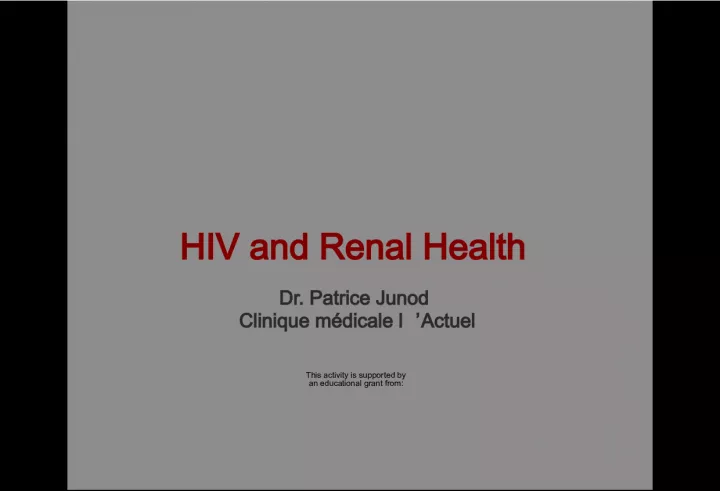
1. HIV and Renal Health Dr. Patrice Junod Clinique mdicale l Actuel This activity is supported by an educational grant from:
2. Content Contributors Principle Content Development Gord Arbess Jean-Guy Baril Mlanie Hamel Brian Conway Chris Fraser Marianne Harris Christine Hughes Patrice Junod Marek Smieja Graham Smith Rachel Therrien Alice Tseng Sharon Walmsley Consultant Linda Robinson Anita Rachlis Ali Zahirieh David Fletcher Program Development
3. This program was developed with consultants through an educational grant from Janssen Inc. The faculty members received financial compensation for developing & presenting this program . Conflict of Interest Declaration
4. Faculty Disclosures Abbvie Gilead Janssen Merck ViiV
5. Discuss factors that can impact renal health in HIV patients List which renal lab tests are the most clinically relevant and how often they should be performed Present a practical tool for the management of declining renal function App ly these learnings using interactive patient case examples Objectives
6. Background: HIV and the Kydney
7. Kidney disease is an important complication of HIV infection in the era of antiretroviral therapy 1 In a retrospective study of 487 consecutive HIV positive patients with normal renal function, the initial prevalence of CKD was 2% 2 After 5 years of follow-up, 6% had progressed to CKD Older age was a multivariate predictor of CKD for this cohort 1 Gupta SK, et al. Clinical Infectious Disease 2005;40:1559-1585. 2 Gupta SK, et al. Clinical Nephrology 2004;61:1-6. Renal Disease in HIV Positive Patients
8. The spectrum of kidney disease in HIV includes: HIV-associated nephropathy Immune complex kidney disease Medication nephrotoxicity Kidney disease related to co-morbid conditions Diabetes, hypertension, and hepatitis virus co-infection Wyatt CM. AJM 2007;120:488-49. Kidney Disease in HIV Positive Patients
9. Age Family History Nephrotoxic medication Diabetes HIV Hyper- tension Hepatitis C Ethnicity CKD Risk = Modifiable = Nonmodifiable Gupta SK, et al. Clinical Infectious Disease 2005;40:1559-1585. Risk Factors for Kidney Disease in the HIV Positive Population
10. Prevalence 3-15% Race and other genetic factors Hypertension Diabetes mellitus Hepatitis C virus infection Decreased CD4 cell count Increased viral load Nephrotoxic Drugs Chronic Kidney Disease in HIV
11. Adapted from: Guo X, Nzerue C. Cleve Clin J Med 2002;69:289-312. TMP/SMX: trimethoprim and sulfamethoxazole Prerenal Tubular Injury Allergic Interstitial Nephritis Thrombotic Microangiopathy Obstructive ACE-I ARBs Direct Renin Inhibitors Amphotericin NSAIDS Cyclosporine Diuretics Interferon Cidofovir Adefovir Tenofovir Didanosine Lamivudine Stavudine Aminoglycosides Amphotericin Cocaine Foscarnet Pentamidine Ketamin Abacavir Indinavir Ritonavir Atazanavir Acyclovir Cephalosporins Penicillins Ciprofloxacin TMP/SMX Rifampin NSAIDs Proton Pump Inhibitors Indinavir Cocaine Cyclosporine Valacyclovir Indinavir Atazanavir Acyclovir Foscarnet Sulfadiazine TMP/SMX Medications and Renal Disease
12. Acute Kidney Injury (AKI) is more common in individuals with HIV infection Chronic Kidney Disease (CKD) is more common in individuals with HIV infection Proteinuria is more common in individuals with HIV infection Proximal tubular dysfunction is more common in individuals with HIV infection HIV & The Kidney: Summary
13. Stage Description GFR (mL/min/1.73m 2 ) I Urinary and/or Structural Abnormality > 89 II Urinary and/or Structural Abnormality 60 - 89 IIIa Mild GFR decline 45 - 59 IIIb Moderate GFR decline 30 - 44 IV Severe GFR decline 15 - 29 V Kidney Failure < 15 ESRD Requiring Renal Replacement Therapy Levey A. KI 2010;80: 17. Classification of CKD
14. Glomerular Filtration Rate (GFR) Proteinuria Proximal Tubular Function Three Important Measures
15. Glomerular Filtration Rate (GFR) Proteinuria Proximal Tubular Function Three Important Measures
16. Gold standard: inulin clearance iothalamate clearance Iohexol Practical serum creatinine 24-hr urine collection for creatinine clearance (cumbersome!) equations, equations, equations How Do We Measure GFR?
17. Serum creatinine Metabolism of creatine in skeletal muscle and from dietary meat intake Production tied to muscle mass Age, weight, sex, amputations, corticosteroid use Modestly influenced by diet Filtered by glomerulus and secreted by proximal tubule Proportionally increased secretion with reduced GFR Creatinine may not increase until up to 50% of GFR is lost Secretion inhibited by drugs including cimetidine, trimethoprim, dapsone, cobicistat Large intra-person and intra-laboratory variation Intra-person variation 720% Poor intra-laboratory calibration particularly affecting higher GFRs Krop JS, et al. Arch Intern Med 1999;159:1777-1783. Coresh J, et al. Am J Kidney Dis 2002;39:920-929. Renal Function Measurement
18. Serum Creatinine 110 mol/L
19. The same serum creatinine represents very different GFRs in these two individuals 40 ml/min 140 ml/min Serum Creatinine 110 mol/L
20. CrCl = weight x (140 age) / (serum Cr x 49)* Estimates CrCl (not GFR) Derived from a study of 249 white Canadian hospitalized veterans who had 2 similar 24-hr urine CrCl measurements Validated for renal dosing of drugs * X 0.8 if female Cockcroft-Gault Equation
21. MDRD GFR (mL/min/1.73m2) = 175 x [serum creatinine(mol/L)/88.4] -1.154 x (Age) -0.203 x (0.742 if female) x (1.21 if African American) Estimates Glomerular Filtration Rate Derived from 1070 individuals with advanced chronic kidney disease 60% male, 88% white, 6% DM MDRD Equation
22. Newest Equation of the three Non-linear based equation More accurate in estimating GFR in those with mild CKD CKD-EPI Levey et al. Ann Intern Med 2009;150: 604-612.
23. Glomerular Filtration Rate (GFR) Proteinuria Proximal Tubular Function Three Important Measures
24. Normal < 150 mg/day of proteinuria < 30 mg/day of albuminuria Quantification strategies: Dipstick Measure ONLY albumin at a CONCENTRATION > 300 mg/L 24-hr urine collection Helpful if patient performs a complete collection Spot urine albumin:creatinine (or protein:creatinine) Can increase sensitivity for detecting proteinuria in a convenient fashion Quantifying Proteinuria
25. A typical man produces roughly 15 mmol of creatinine/day A typical woman produces roughly 10 mmol of creatinine/day The protein:creatinine (PCR) or albumin:creatinine (ACR) tell you how much protein/albumin is present in the urine per mmol of Cr Thus multiplying the ACR by 10 in woman and by 15 in men will give you an estimate of that individuals 24hr excretion of albumin (the exact same applies to PCR) Practical Point
26. A marker of increased risk of CV events Increased risk of CKD progression (notably when > 1g/day protein or 200mg/day albumin) Implications of Proteinuria
27. Glomerular Filtration Rate (GFR) Proteinuria Proximal Tubular Function Three Important Measures
28. Reabsorption Water Electrolytes Bicarbonate Glucose Filtered proteins Secretion Organic Anions/Cations Drugs Metabolic Byproducts Creatinine Ernst M, Moser M. N Engl J Med 2009;361:2153-2164. Tubular Functions
29. Ernst M, Moser M. N Engl J Med 2009;361:2153-2164. Proximal Tubular Function Protein Reabsorption Phosphate Reabsorption Glucose Reabsorption Amino Acid Reabsorption Creatinine Secretion Bicarbonate reabsorption
30. Some Evidence of Proximal Tubular Injury Urine: glucosuria in absence of diabetes Non-albumin based proteinuria measure both albuminuria & proteinuria high urinary 2-microglobulin excretion Evidence of ATN (hemegranular casts) Serum: non-anion gap metabolic acidosis, creatinine rise Hypophosphatemia & high urinary phosphate excretion Calculate the Fractional Excretion of Phosphate* (Urinary PO4/Ur Cr) / (Serum PO4/Serum Cr) Abnomal = greater than 10% in setting of hypophosphatemia What Are You Looking For?
31. Aquitaine Cohort 399 patients in a cross sectional analysis Overall prevalence of PRTD was high at 6.5 % 29.6 % stage 1 or 2 kidney disease 5.3 % stage 3 to 5 kidney disease F-A Dauchy et al. Kidney International 2011;80:302-309. Increased risk of abnormal proximal renal tubular function with HIV infection and antiretroviral therapy
32. Multivariate Analysis showed significant independent associations Age (OR 1.28 per 5 year increase) TDF (OR 1.23 per year) ATZ (OR 1.28) Primary tubular abnormalities can be missed even when severe and can lead to decline in GFR Early screening is necessary to avoid them Increased risk of abnormal proximal renal tubular function with HIV infection and antiretroviral therapy F-A Dauchy et al. Kidney International 2011;80:302-309.
33. Guidelines
34. IDSA Guidelines: Evaluating and Monitoring CKD in HIV All patients at the time of HIV diagnosis should be assessed for existing kidney disease Calculated estimate of renal function and Screening for proteinuria Dipstick, protein/creat ratio or albumin/creat ratio? If there is no evidence of kidney disease at initial evaluation, patients at high risk for the development of proteinuric renal disease should undergo annual screening African American persons CD4+ cell counts <200 mL or HIV RNA levels >4000 copies/mL Diabetes mellitus Hypertension Hepatitis C virus coinfection Patients without risk factors for kidney disease should be followed clinically and reassessed based on the occurrence of signs and symptoms or as clinical events dictate Gupta SK et al. Clin Infect Dis 2005;40:1559-1585.
35. IDSA Initial Evaluation Recommendations Obtain baseline GFR: All patients at the time of HIV diagnosis should be assessed for existing kidney disease with a screening urinalysis for proteinuria and a calculated estimate of renal function Annual screening: If there is no evidence of proteinuria at initial evaluation, patients at high risk for the development of proteinuric renal disease should undergo annual screening Renal function should be estimated on a yearly basis to assess for changes over time When to consider a nephrology consult: Additional evaluations and referral to a nephrologist are recommended for patients with proteinuria of grade 1+ by dipstick analysis or GFR<60 mL/min per 1.73m 2 Gupta SK, et al. Clinical Infectious Disease 2005;40:1559-1585.
36. DHHS Recommendations Panel on Antiretroviral Guidelines for Adults and Adolescents. Guidelines for the use of antiretroviral agents in HIV-1-infected adults and adolescents. Department of Health and Human Services. Available at http://aidsinfo.nih.gov/ContentFiles/Adultand Entry info care Follow-up before ART ART Initiation or modification Follow-up 2-8 weeks post- ART initiation or modification Every 3-6 months Every 6 months Every 12 months Treatme nt failure Clinically indicated ALT, AST, T bilirubin Every 6-12 months CBC with differential Every 3-6 months If on ZDV Fasting lipid profile If normal annually Consider 4-8 weeks after starting new ART regimen that affects lipids If abnormal at last measurement If abnormal at last measurem ent Fasting glucose or hemoglobin A1C If normal annually If abnormal at last measurement If abnormal at last measurement Urinalysis If on TDF Pregnancy test If startting EFV Table 3. Laboratory Monitoring Schedule for Patients Before and After Initiation of Antiretroviral Therapy
37. Kidney Stones Chronic Kidney Disease (CKD) Emerging Evidence
38. Renal stones are risk factor for chronic kidney disease (CKD) Urolithiasis well-known side effect of indinavir Considered to be drug crystallization in urine Urolithiasis also associated with atazanavir Probably similar etiology Hamada et al. Clin Infect Dis, 2012 Renal Stones
39. Cohort analysis of 1240 patients ATV/r (n=465) or other protease inhibitors (n=775) Renal stones developed in 31 patients on ATV/r (6.7%) and 4 patients (0.52%) on other PIs Risk was 10 times higher in ATV/r group Patients on ATV/r had lower eGFR Lower eGFR associated with renal stones Hamada et al. Clin Infect Dis, 2012 ATV & Renal Stones: Hamada et al.
40. Event rate remained significantly higher in the ATV cohort after adjusting for prior ATV and IDV exposure ATV/r patients who developed renal stones had significantly higher bilirubin levels vs. ATV/r patients who did not develop stones At study baseline, 42% of ATV/r patients who developed renal stones had chronic renal impairment vs. 4.5% of ATV/r patients who did not develop stones ATV (n = 1,206) EFV / DRV / LPV combined cohort (n=4,449) p value No. of patients with kidney stones 24 24 Prevalence of kidney stones per 1,000 patients (95% CI) 20 (13 - 30) 5.4 (3.2 7.6) < 0.001 Event rate per 1,000 pt-yrs of exposure, n (95% CI) 7.3 (4.7 - 10.8) 1.9 (1.2 - 2.8) < 0.001 Rockwood N, et al. 17 e confrence annuelle de la BHIVA, Bournemouth, 2011, rsum O4. ATV & Renal Stones: Rockwood et al.
41. *Adjusted for gender, age at start of HAART, ethnicity, baseline eGFR, baseline CD4 cell count, baseline viral load, HBsAg, prior exposure to TDF and IDV and total duration of TDF exposure Rockwood N, et al. J Antivir Antiretrovir 2012;4: 21-25. Hazard ratio* (95% CI) p value LPV/r 1.69 (1.1 - 2.6) 0.017 ATV/r 1.52 (1.14 - 2.03) 0.004 DRV/r 1.31 (0.94 1.81) 0.108 EFV 1.00 Au cours des 12 premiers mois, 49 % des sujets ayant dvelopp une insuffisance rnale s taient rtablis (TFGe > 60 ml/min/1,73 m 2 ). Rockwood et al., J Antivir Antiretrovir 2012, 4:2 Renal Impairment PIs vs EFV
42. Daar et al. Ann Intern Med , 2011 n 338 287 377 330 360 327 394 352 Median Change in Calculated Creatinine from Baseline (mL/min) ** * ATV/r EFV EFV ATV/r +ABC/3TC +TDF/FTC Median Creatinine Clearance: ATV/r vs. EFV * p = 0,001 p/r at ATV/r ** p < 0,001 p/r at ATV/r Week 48 Week 96 ACTG 5202: Creatinine Clearance
43. Medication Annual Increased Risk Atazanavir + Tenofovir 41 % Atazanavir 22 % Tenofovir 16 % Indinavir 11 % Lopinavir/r 8 % Adapt de Mocroft et al. AIDS, 2010 N = 6,843 Mean follow up was 3.7 years Incidence of CKD with Each Additional Year of Exposure Chronic Kidney Disease & ARV Exposure
44. Cohort of 49,734 First analysis to focus on patients with normal renal function at baseline (n=22,603) eGFR > 90 ml/min/1.73m 2 Followed to confirmed: eGFR < 70 ml/min/1.73m 2 Or eGFR < 60 ml/min/1.73m 2 Or last available eGFR Ryom et al. Prsentation d affiche, CROI, 2012 ARVs & Renal Impairment: The D:A:D Cohort
45. N=22,603 4.5 year follow up 468 (2.1%) patients progressed to eGFR < 70 incidence rate 4.78/1000 patient years 131 (0.6%) patients progressed to CKD incidence rate 1.33/1000 patient years Equals an annual decline of at least 4-5 ml/min Ryom et al. Prsentation d affiche, CROI, 2012 CKD=Chronic Kidney Disease D:A:D Cohort: Results
46. Ryom et al. Poster presentation , CROI, 2012 ARVs Exposure Rates of ceGFR <70 from eGFR > 90 (adjusted analysis)
47. Medication Adjusted Hazard Ratio (95% CI) P Value Non PI 1.00 Tenofovir 1.16 0.177 Lopinavir 1.32 0.024 Atazanavir 1.46 < 0.001 Adapt de Hosein et al. Prsentation d affiche, IAS, 2011 N = 965 Time to Impaired eGFR Canadian Observational Cohort (CANOC) Collaboration
48. EuroSIDA Study: Risk for Chronic Kidney Disease Analysis of patients with 3 creatinine measurements + body weight 6,842 patients with 21,482 person-years of follow-up Definition of CKD (eGFR by Cockcroft-Gault) If baseline eGFR 60 mL/min/1.73 m 2 , fall to <60 If baseline eGFR <60 mL/min/1.73 m 2 , fall by 25% 225 (3.3%) progressed to CKD Risk factors for CKD on TDF: age, HTN, HCV, lower eGFR, lower CD4+ count Cumulative Exposure to ARVs and Risk of CKD Kirk O, et al. 17th CROI; San Francisco, CA; February 16-19, 2010. Abst. 107LB. Univariable Multivariable IRR/year 95% CI P-value IRR/year 95% CI P-value Tenofovir 1.32 1.21-1.41 <0.0001 1.16 1.06-1.25 <0.0001 Indinavir 1.18 1.13-1.24 <0.0001 1.12 1.06-1.18 <0.0001 Atazanavir 1.48 1.35-1.62 <0.0001 1.21 1.09-1.34 0.0003 Lopinavir/r 1.15 1.07-1.23 <0.0001 1.08 1.01-1.16 0.030
49. EuroSIDA STUDY: Crude Incidence Rate of CKD and Increasing Exposure to ARVs Kirk, CROI 2010; 107LB. CKD, confirmed (persisting for >3 months) decrease in eGFR 60 mL/min/1.73m 2 if eGFR at baseline >60 mL/min/1.73m 2 or confirmed 25% decrease in eGFR if baseline eGFR60 mL/min/1.73m 2 Incidence per 100 PYFU (95% CI) Years of Exposure to ARV N with CKD Not 0-1 1-2 2-3 >3 started Not 0-1 1-2 2-3 >3 86 21 34 29 55 67 31 35 25 67 Not 0-1 1-2 2-3 >3 127 20 19 11 48 Not 0-1 1-2 2-3 >3 143 23 20 18 21 started started started Tenofovir Indinavir Atazanavir Lopinavir/r 10 1 .01
50. 1- Algorithm Nephropathy Advisory Committee on the clinical management of people living with HIV 2- HIV and Renal Health Management tool National Development Committee Supported by Janssen Algorithm
51. Nephropathy Advisory Committee on the Clinical Management of Persons Living with HIV PERIODIC HEALTH EXAMINATION OF ADULTS LIVING WITH HIV (HUMAN IMMUNODEFICIENCY VIRUS)
52. Screening schedule based on risk factors for kidney disease (EACS 2011) Untreated HIV+ patients Treated HIV+ patients Without TDF With TDF Assessment of risk factors for CKD* Annual Annual 612 months Urinalysis or urine dipstick Annual Annual 6 months if GFR < 60 3-6 months eGFR 6-12 months 3-6 months 3-6 months Phosphorus As needed As needed Optional 3-6 months * Risk factors for CKD: Diabetes, hypertension, CVD, viral hepatitis, concomitant nephrotoxic drugs, family history of CKD, black African ethnicity Advisory Committee on the Clinical Management of Persons Living with HIV Screening for Kidney Problems
53. GFR using CKD-EPI or MDRD GFR using CKD-EPI or MDRD ACR and MAU ACR and MAU Refer to proteinuria algorithm (next page) Refer to proteinuria algorithm (next page) Referral to nephrologist or internist Referral to nephrologist or internist < 60 cc/min* < 60 cc/min* < 30 cc/min* < 30 cc/min* CaPO4 CaPO4 Renal ultrasound Renal ultrasound > 60 and < 90 cc/min > 60 and < 90 cc/min Increase in Cr > 20% for > 3 months** Increase in Cr > 20% for > 3 months** Repeat CKD-EPI or MDRD calculation Repeat CKD-EPI or MDRD calculation Refer to algorithms (next pages) Refer to algorithms (next pages) GFR < 90 GFR < 90 Glucose+ Protein+ HypoPO4 Glucose+ Protein+ HypoPO4 GFR > 90 GFR > 90 Regular follow-up Regular follow-up Follow up every 3 months Follow up every 3 months * If GFR < 50 cc/min: consider adjusting the dose of certain ARV and concomitant medications ** Test for tubulopathy if GFR declines > 10 cc/min while on tenofovir
54. GFR using CKD-EPI or MDRD GFR using CKD-EPI or MDRD ACR and MAU ACR and MAU Refer to proteinuria algorithm (next page) Refer to proteinuria algorithm (next page) Referral to nephrologist or internist Referral to nephrologist or internist < 60 cc/min* < 60 cc/min* < 30 cc/min* < 30 cc/min* CaPO4 CaPO4 Renal ultrasound Renal ultrasound > 60 and < 90 cc/min > 60 and < 90 cc/min Increase in Cr > 20% for > 3 months** Increase in Cr > 20% for > 3 months** Repeat CKD-EPI or MDRD calculation Repeat CKD-EPI or MDRD calculation Refer to algorithms (next pages) Refer to algorithms (next pages) GFR < 90 GFR < 90 Glucose+ Protein+ HypoPO4 Glucose+ Protein+ HypoPO4 GFR > 90 GFR > 90 Regular follow-up Regular follow-up Follow up every 3 months Follow up every 3 months * If GFR < 50 cc/min: consider adjusting the dose of certain ARV and concomitant medications ** Test for tubulopathy if GFR declines > 10 cc/min while on tenofovir
55. * If GFR < 50 cc/min: consider adjusting the dose of certain ARV and concomitant medications ** Test for tubulopathy if GFR declines > 10 cc/min while on tenofovir > 60 and < 90 cc/min > 60 and < 90 cc/min Increase in Cr > 20% for > 3 months** Increase in Cr > 20% for > 3 months** Repeat CKD-EPI or MDRD calculation Repeat CKD-EPI or MDRD calculation Refer to algorithms (next pages) Refer to algorithms (next pages) GFR < 90 GFR < 90 Glucose+ Protein+ HypoPO4 Glucose+ Protein+ HypoPO4 GFR > 90 GFR > 90 Regular follow-up Regular follow-up Follow up every 3 months Follow up every 3 months GFR using CKD-EPI or MDRD GFR using CKD-EPI or MDRD
56. GFR using CKD-EPI or MDRD GFR using CKD-EPI or MDRD ACR and MAU ACR and MAU Refer to proteinuria algorithm (next page) Refer to proteinuria algorithm (next page) Referral to nephrologist or internist Referral to nephrologist or internist < 60 cc/min* < 60 cc/min* < 30 cc/min* < 30 cc/min* CaPO4 CaPO4 Renal ultrasound Renal ultrasound > 60 and < 90 cc/min > 60 and < 90 cc/min Increase in Cr > 20% for > 3 months** Increase in Cr > 20% for > 3 months** Repeat CKD-EPI or MDRD calculation Repeat CKD-EPI or MDRD calculation Refer to algorithms (next pages) Refer to algorithms (next pages) GFR < 90 GFR < 90 Glucose+ Protein+ HypoPO4 Glucose+ Protein+ HypoPO4 GFR > 90 GFR > 90 Regular follow-up Regular follow-up Follow up every 3 months Follow up every 3 months * If GFR < 50 cc/min: consider adjusting the dose of certain ARV and concomitant medications ** Test for tubulopathy if GFR declines > 10 cc/min while on tenofovir
57. GFR using CKD-EPI or MDRD GFR using CKD-EPI or MDRD ACR and MAU ACR and MAU Refer to proteinuria algorithm (next page) Refer to proteinuria algorithm (next page) Referral to nephrologist or internist Referral to nephrologist or internist < 60 cc/min* < 60 cc/min* < 30 cc/min* < 30 cc/min* CaPO4 CaPO4 Renal ultrasound Renal ultrasound * If GFR < 50 cc/min: consider adjusting the dose of certain ARV and concomitant medications ** Test for tubulopathy if GFR declines > 10 cc/min while on tenofovir
58. Urinalysis or urine dipstick Urinalysis or urine dipstick Glucose > 0 Glucose > 0 Glycosuri a DB + Glycosuri a DB + Glycosuri a DB Glycosuri a DB DB follow-up DB follow-up Fasting glucose + Rule out diabetes Fasting glucose + Rule out diabetes Repeat 1x Repeat 1x Glycosuri a DB Glycosuri a DB Referral to nephrologist or internist Referral to nephrologist or internist ACR 0.05 g/mmol and MAU < 2.1 mg/mmol ACR 0.05 g/mmol and MAU < 2.1 mg/mmol Normal Normal - Renal ultrasound - Ascertain the risk factors - Referral to nephrologist or internist, or to urologist for isolated hematuria - Renal ultrasound - Ascertain the risk factors - Referral to nephrologist or internist, or to urologist for isolated hematuria Protein 1 + or 0.25 g/L Protein 1 + or 0.25 g/L Repeat at next appt. Repeat at next appt. Protein < 1+ or 0.25 g/L Protein < 1+ or 0.25 g/L Protein 1+ or 0.25 g/L Protein 1+ or 0.25 g/L Normal Normal ACR and MAU ACR and MAU ACR > 0.05 g/mmol or MAU > 2.1 mg/mmol or hematuria (> 2 RBC/HPF) ACR > 0.05 g/mmol or MAU > 2.1 mg/mmol or hematuria (> 2 RBC/HPF)
59. Urinalysis or urine dipstick Urinalysis or urine dipstick Glucose > 0 Glucose > 0 Glycosuri a DB + Glycosuri a DB + Glycosuri a DB Glycosuri a DB DB follow-up DB follow-up Fasting glucose + Rule out diabetes Fasting glucose + Rule out diabetes Repeat 1x Repeat 1x Glycosuri a DB Glycosuri a DB Referral to nephrologist or internist Referral to nephrologist or internist ACR 0.05 g/mmol and MAU < 2.1 mg/mmol ACR 0.05 g/mmol and MAU < 2.1 mg/mmol Normal Normal - Renal ultrasound - Ascertain the risk factors - Referral to nephrologist or internist, or to urologist for isolated hematuria - Renal ultrasound - Ascertain the risk factors - Referral to nephrologist or internist, or to urologist for isolated hematuria Protein 1 + or 0.25 g/L Protein 1 + or 0.25 g/L Repeat at next appt. Repeat at next appt. Protein < 1+ or 0.25 g/L Protein < 1+ or 0.25 g/L Protein 1+ or 0.25 g/L Protein 1+ or 0.25 g/L Normal Normal ACR and MAU ACR and MAU ACR > 0.05 g/mmol or MAU > 2.1 mg/mmol or hematuria (> 2 RBC/HPF) ACR > 0.05 g/mmol or MAU > 2.1 mg/mmol or hematuria (> 2 RBC/HPF)
60. Urinalysis or urine dipstick Urinalysis or urine dipstick Glucose > 0 Glucose > 0 Glycosuri a DB + Glycosuri a DB + Glycosuri a DB Glycosuri a DB DB follow-up DB follow-up Fasting glucose + Rule out diabetes Fasting glucose + Rule out diabetes Repeat 1x Repeat 1x Glycosuri a DB Glycosuri a DB Referral to nephrologist or internist Referral to nephrologist or internist
61. Urinalysis or urine dipstick Urinalysis or urine dipstick Glucose > 0 Glucose > 0 Glycosuri a DB + Glycosuri a DB + Glycosuri a DB Glycosuri a DB DB follow-up DB follow-up Fasting glucose + Rule out diabetes Fasting glucose + Rule out diabetes Repeat 1x Repeat 1x Glycosuri a DB Glycosuri a DB Referral to nephrologist or internist Referral to nephrologist or internist ACR 0.05 g/mmol and MAU < 2.1 mg/mmol ACR 0.05 g/mmol and MAU < 2.1 mg/mmol Normal Normal - Renal ultrasound - Ascertain the risk factors - Referral to nephrologist or internist, or to urologist for isolated hematuria - Renal ultrasound - Ascertain the risk factors - Referral to nephrologist or internist, or to urologist for isolated hematuria Protein 1 + or 0.25 g/L Protein 1 + or 0.25 g/L Repeat at next appt. Repeat at next appt. Protein < 1+ or 0.25 g/L Protein < 1+ or 0.25 g/L Protein 1+ or 0.25 g/L Protein 1+ or 0.25 g/L Normal Normal ACR and MAU ACR and MAU ACR > 0.05 g/mmol or MAU > 2.1 mg/mmol or hematuria (> 2 RBC/HPF) ACR > 0.05 g/mmol or MAU > 2.1 mg/mmol or hematuria (> 2 RBC/HPF)
62. Urinalysis or urine dipstick Urinalysis or urine dipstick ACR 0.05 g/mmol and MAU < 2.1 mg/mmol ACR 0.05 g/mmol and MAU < 2.1 mg/mmol Normal Normal - Renal ultrasound - Ascertain the risk factors - Referral to nephrologist or internist, or to urologist for isolated hematuria - Renal ultrasound - Ascertain the risk factors - Referral to nephrologist or internist, or to urologist for isolated hematuria Protein 1 + or 0.25 g/L Protein 1 + or 0.25 g/L Repeat at next appt. Repeat at next appt. Protein < 1+ or 0.25 g/L Protein < 1+ or 0.25 g/L Protein 1+ or 0.25 g/L Protein 1+ or 0.25 g/L Normal Normal ACR and MAU ACR and MAU ACR > 0.05 g/mmol or MAU > 2.1 mg/mmol or hematuria (> 2 RBC/HPF) ACR > 0.05 g/mmol or MAU > 2.1 mg/mmol or hematuria (> 2 RBC/HPF)
63. Serum phosphorus Serum phosphorus < normal levels < normal levels Repeat and if < normal levels Repeat and if < normal levels PTH assay PTH assay 25-OH Vit D 25-OH Vit D Albumin- corrected Ca Albumin- corrected Ca < 50: deficiency < 75: insufficiency < 50: deficiency < 75: insufficiency > 75 > 75 Vit D Rx Vit D Rx Normal Normal Abnorma l Abnorma l Normal Normal Referral to nephrologist or internist Referral to nephrologist or internist Urinary fractional excretion of phosphorus if available (if > 20% or > 10% and hypophosphatemia: referral to a specialist Urinary fractional excretion of phosphorus if available (if > 20% or > 10% and hypophosphatemia: referral to a specialist Abnorma l Abnorma l Normal Normal Referral to nephrologist or internist Referral to nephrologist or internist 0.65 normal level 0.65 normal level 0.32 0.65 mmol/L 0.32 0.65 mmol/L < 0.32 mmol/L < 0.32 mmol/L Repeat in 3 months Repeat in 3 months Repeat in 1 month Repeat in 1 month Treat immediately Referral to nephrologist Treat immediately Referral to nephrologist
64. Serum phosphorus Serum phosphorus < normal levels < normal levels Repeat and if < normal levels Repeat and if < normal levels PTH assay PTH assay 25-OH Vit D 25-OH Vit D Albumin- corrected Ca Albumin- corrected Ca < 50: deficiency < 75: insufficiency < 50: deficiency < 75: insufficiency > 75 > 75 Vit D Rx Vit D Rx Normal Normal Abnorma l Abnorma l Normal Normal Referral to nephrologist or internist Referral to nephrologist or internist Urinary fractional excretion of phosphorus if available (if > 20% or > 10% and hypophosphatemia: referral to a specialist Urinary fractional excretion of phosphorus if available (if > 20% or > 10% and hypophosphatemia: referral to a specialist Abnorma l Abnorma l Normal Normal Referral to nephrologist or internist Referral to nephrologist or internist 0.65 normal level 0.65 normal level 0.32 0.65 mmol/L 0.32 0.65 mmol/L < 0.32 mmol/L < 0.32 mmol/L Repeat in 3 months Repeat in 3 months Repeat in 1 month Repeat in 1 month Treat immediately Referral to nephrologist Treat immediately Referral to nephrologist
65. Serum phosphorus Serum phosphorus < normal levels < normal levels Repeat and if < normal levels Repeat and if < normal levels Urinary fractional excretion of phosphorus if available (if > 20% or > 10% and hypophosphatemia: referral to a specialist Urinary fractional excretion of phosphorus if available (if > 20% or > 10% and hypophosphatemia: referral to a specialist 0.65 normal level 0.65 normal level 0.32 0.65 mmol/L 0.32 0.65 mmol/L < 0.32 mmol/L < 0.32 mmol/L Repeat in 3 months Repeat in 3 months Repeat in 1 month Repeat in 1 month Treat immediately Referral to nephrologist Treat immediately Referral to nephrologist
66. Serum phosphorus Serum phosphorus < normal levels < normal levels Repeat and if < normal levels Repeat and if < normal levels PTH assay PTH assay 25-OH Vit D 25-OH Vit D Albumin- corrected Ca Albumin- corrected Ca < 50: deficiency < 75: insufficiency < 50: deficiency < 75: insufficiency > 75 > 75 Vit D Rx Vit D Rx Normal Normal Abnorma l Abnorma l Normal Normal Referral to nephrologist or internist Referral to nephrologist or internist Urinary fractional excretion of phosphorus if available (if > 20% or > 10% and hypophosphatemia: referral to a specialist Urinary fractional excretion of phosphorus if available (if > 20% or > 10% and hypophosphatemia: referral to a specialist Abnorma l Abnorma l Normal Normal Referral to nephrologist or internist Referral to nephrologist or internist 0.65 normal level 0.65 normal level 0.32 0.65 mmol/L 0.32 0.65 mmol/L < 0.32 mmol/L < 0.32 mmol/L Repeat in 3 months Repeat in 3 months Repeat in 1 month Repeat in 1 month Treat immediately Referral to nephrologist Treat immediately Referral to nephrologist
67. Serum phosphorus Serum phosphorus < normal levels < normal levels Repeat and if < normal levels Repeat and if < normal levels PTH assay PTH assay 25-OH Vit D 25-OH Vit D Albumin- corrected Ca Albumin- corrected Ca < 50: deficiency < 75: insufficiency < 50: deficiency < 75: insufficiency > 75 > 75 Vit D Rx Vit D Rx Normal Normal Abnorma l Abnorma l Normal Normal Referral to nephrologist or internist Referral to nephrologist or internist Urinary fractional excretion of phosphorus if available (if > 20% or > 10% and hypophosphatemia: referral to a specialist Urinary fractional excretion of phosphorus if available (if > 20% or > 10% and hypophosphatemia: referral to a specialist Abnorma l Abnorma l Normal Normal Referral to nephrologist or internist Referral to nephrologist or internist
68. Algorithm
69. Algorithm
70. Algorithm
71. Algorithm
72. Algorithm
73. Case Study Aging Woman with longstanding HIV and multiple comorbidities Dr. Gord Arbess
74. 62 year old woman 62 year old woman From Jamaica From Jamaica HIV + since 1996, heterosexual transmission HIV + since 1996, heterosexual transmission Nadir CD4 108, VL > 500,000 Nadir CD4 108, VL > 500,000 Intermittent adherence Intermittent adherence Multiple ARV Regimens due to intolerance/resistance (AZT, 3TC, ddI, d4T, Nelfinavir, Amprenavir, LPV, EFV, Indinavir, Tenofovir, RTV) Multiple ARV Regimens due to intolerance/resistance (AZT, 3TC, ddI, d4T, Nelfinavir, Amprenavir, LPV, EFV, Indinavir, Tenofovir, RTV) Hx ABC/3TC HSR Hx ABC/3TC HSR Background Information
75. Obese Obese Hypertension Hypertension NIDDM (Gastroparesis-intermittent vomiting) NIDDM (Gastroparesis-intermittent vomiting) Sleep Apnea-CPAP Sleep Apnea-CPAP Angina? Angina? Severe Osteoarthritis Knees Severe Osteoarthritis Knees Hypothyroid Hypothyroid Hyperlipidemia Hyperlipidemia Major Depression Major Depression Multiple Co-Morbidities
76. Present HIV Regimen started June 2012 Darunavir 800 mg/d Ritonavir 100 mg/d Raltegravir 400 mg bid Etravirine 400 mg/d HIV Medications
77. Lisinopril Lisinopril Atorvastatin Atorvastatin Ibuprofen Ibuprofen Metformin Metformin Cipralex Cipralex Zofran Zofran Eltroxin Eltroxin Other Medications
78. You notice Serum Cr is 158 (eGFR 48) on routine BW in August 2012 Routine Bloodwork
79. What Would You Do?
80. GFR using CKD-EPI or MDRD GFR using CKD-EPI or MDRD ACR and MAU ACR and MAU Refer to proteinuria algorithm (next page) Refer to proteinuria algorithm (next page) Referral to nephrologist or internist Referral to nephrologist or internist < 60 cc/min* < 60 cc/min* < 30 cc/min* < 30 cc/min* CaPO4 CaPO4 Renal ultrasound Renal ultrasound * If GFR < 50 cc/min: consider adjusting the dose of certain ARV and concomitant medications ** Test for tubulopathy if GFR declines > 10 cc/min while on tenofovir
81. Algorithm
82. Urinalysis ACR Serum Cr (eGFR) Electrolytes, Bicarb, albumin Urine for Protein, Cr Renal Ultrasound Other? Biopsy? Investigations to assess Renal Function
83. VL < 40 CD 4 843 Hgb 108 BS 7.3 Hga1c 0.061 ACR 1.1 Trace Protein, no blood, no glucose, 10-15 White cells/hpf, occ red cells/hpf, hyaline casts with some cells Spot urine 0.1 g/L protein, 7.8 mmol/L Cr Cr 118-160 range (eGFR 48-54 range) over number of years Normal electrolytes, normal albumin, normal Bicarb Normal renal Ultrasound (small-sized kidneys) Results
84. What Would You Do?
85. Urinalysis or urine dipstick Urinalysis or urine dipstick Glucose > 0 Glucose > 0 Glycosuri a DB + Glycosuri a DB + Glycosuri a DB Glycosuri a DB DB follow-up DB follow-up Fasting glucose + Rule out diabetes Fasting glucose + Rule out diabetes Repeat 1x Repeat 1x Glycosuri a DB Glycosuri a DB Referral to nephrologist or internist Referral to nephrologist or internist ACR 0.05 g/mmol and MAU < 2.1 mg/mmol ACR 0.05 g/mmol and MAU < 2.1 mg/mmol Normal Normal - Renal ultrasound - Ascertain the risk factors - Referral to nephrologist or internist, or to urologist for isolated hematuria - Renal ultrasound - Ascertain the risk factors - Referral to nephrologist or internist, or to urologist for isolated hematuria Protein 1 + or 0.25 g/L Protein 1 + or 0.25 g/L Repeat at next appt. Repeat at next appt. Protein < 1+ or 0.25 g/L Protein < 1+ or 0.25 g/L Protein 1+ or 0.25 g/L Protein 1+ or 0.25 g/L Normal Normal ACR and MAU ACR and MAU ACR > 0.05 g/mmol or MAU > 2.1 mg/mmol or hematuria (> 2 RBC/HPF) ACR > 0.05 g/mmol or MAU > 2.1 mg/mmol or hematuria (> 2 RBC/HPF)
86. Algorithm
87. What do you think could be accounting for Cr elevation?
88. HIVAN? HIVAN? IgA Nephropathy? IgA Nephropathy? Medication-related? Medication-related? Hypertension? Hypertension? NIDDM? NIDDM? Pre-renal component/volume contraction? Pre-renal component/volume contraction? Other? Other? Etiology
89. How would you manage this patient?
90. Do you d/c metformin? Do you d/c NSAIDs? Do you d/c statin? Do you Need to dose Adjust ARVs? Should you Change ARVs? Do you Hold Ace Inhibitor? Do you ensure BP/BS well controlled? Do Nothing? Management Options?
91. BP well controlled Hga1c 0.062, therefore Metformin stopped Asked not to take any NSAIDS ARV regimen continued at same doses Continued same dose of statin, ACEi Cr monitored closely in range of 118-130 (eGFR 55-60 range) Follow Up