Chromosome Structure and Function

Learn about the organization and role of chromosomes in genetic inheritance and cell division. Understand the importance of DNA packaging and how it affects gene expression.
- Uploaded on | 1 Views
-
 byron
byron
About Chromosome Structure and Function
PowerPoint presentation about 'Chromosome Structure and Function'. This presentation describes the topic on Learn about the organization and role of chromosomes in genetic inheritance and cell division. Understand the importance of DNA packaging and how it affects gene expression.. The key topics included in this slideshow are . Download this presentation absolutely free.
Presentation Transcript
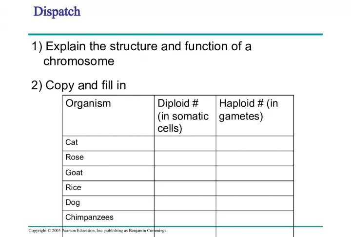
1. Copyright © 2005 Pearson Education, Inc. publishing as Benjamin Cummings Dispatch 1) Explain the structure and function of a chromosome 2) Copy and fill in Organism Diploid # (in somatic cells) Haploid # (in gametes) Cat Rose Goat Rice Dog Chimpanzees
2. Copyright © 2005 Pearson Education, Inc. publishing as Benjamin Cummings PowerPoint Lectures for Biology, Seventh Edition Neil Campbell and Jane Reece Lectures by Chris Romero Chapter 12 Chapter 12 The Cell Cycle
3. Copyright © 2005 Pearson Education, Inc. publishing as Benjamin Cummings • http://highered.mcgraw- hill.com/sites/0072495855/student_view0/chapter2 /animation__how_the_cell_cycle_works.html
4. Copyright © 2005 Pearson Education, Inc. publishing as Benjamin Cummings Mitosis in Action • Spindle=_________ • Spindle=_________ • Spindle=_________ • Spindle=_________ • Nucleus=_________ • Cell Membrane=______ • Chromosome=______ • Chromosome=______
5. Copyright © 2005 Pearson Education, Inc. publishing as Benjamin Cummings • In unicellular organisms, division of one cell reproduces the entire organism • Multicellular organisms depend on cell division for: – Development from a fertilized cell – Growth – Repair
6. Copyright © 2005 Pearson Education, Inc. publishing as Benjamin Cummings Concept 12.1: Cell division results in genetically identical daughter cells • Cells duplicate their genetic material before they divide, ensuring that each daughter cell receives an exact copy of the genetic material, DNA • A dividing cell duplicates its DNA, allocates the two copies to opposite ends of the cell, and only then splits into daughter cells
7. Copyright © 2005 Pearson Education, Inc. publishing as Benjamin Cummings Cellular Organization of the Genetic Material • A cell’s endowment of DNA (its genetic information) is called its genome • DNA molecules in a cell are packaged into chromosomes
8. Copyright © 2005 Pearson Education, Inc. publishing as Benjamin Cummings • Every eukaryotic species has a characteristic number of chromosomes in each cell nucleus • Somatic (nonreproductive) cells have two sets of chromosomes • Gametes (reproductive cells: sperm and eggs) have half as many chromosomes as somatic cells • Eukaryotic chromosomes consist of chromatin , a complex of DNA and protein that condenses during cell division
9. Copyright © 2005 Pearson Education, Inc. publishing as Benjamin Cummings Distribution of Chromosomes During Cell Division • In preparation for cell division, DNA is replicated and the chromosomes condense • Each duplicated chromosome has two sister chromatids, which separate during cell division • The centromere is the narrow “waist” of the duplicated chromosome, where the two chromatids are most closely attached
10. LE 12-4 LE 12-4 Chromosome duplication (including DNA synthesis) 0.5 µm Centromere Sister chromatids Separation of sister chromatids Centromeres Sister chromatids
11. Copyright © 2005 Pearson Education, Inc. publishing as Benjamin Cummings • Eukaryotic cell division consists of: – Mitosis, the division of the nucleus – Cytokinesis, the division of the cytoplasm • Gametes are produced by a variation of cell division called meiosis • Meiosis yields nonidentical daughter cells that have only one set of chromosomes, half as many as the parent cell
12. Copyright © 2005 Pearson Education, Inc. publishing as Benjamin Cummings Phases of the Cell Cycle • The cell cycle consists of – Mitotic (M) phase (mitosis and cytokinesis) – Interphase (cell growth and copying of chromosomes in preparation for cell division) • Interphase (about 90% of the cell cycle) can be divided into subphases: – G 1 phase (“first gap”) – S phase (“synthesis”) – G 2 phase (“second gap”)
13. LE 12-5 LE 12-5 G 1 G 2 S (DNA synthesis) INTERPHASE Cytokinesis MITOTIC (M) PHASE Mitosis
14. Copyright © 2005 Pearson Education, Inc. publishing as Benjamin Cummings • http://bio.rutgers.edu/~gb101/lab2_mitosis/section 2_frames.html
15. Copyright © 2005 Pearson Education, Inc. publishing as Benjamin Cummings The Mitotic Spindle: A Closer Look • The mitotic spindle is an apparatus of microtubules that controls chromosome movement during mitosis • Assembly of spindle microtubules begins in the centrosome, the microtubule organizing center • The centrosome replicates, forming two centrosomes that migrate to opposite ends of the cell, as spindle microtubules grow out from them • An aster (a radial array of short microtubules) extends from each centrosome
16. LE 12-7 LE 12-7 Microtubules Chromosomes Sister chromatids Aster Centrosome Metaphase plate Kineto- chores Kinetochore microtubules 0.5 µm Overlapping nonkinetochore microtubules 1 µm Centrosome
17. Copyright © 2005 Pearson Education, Inc. publishing as Benjamin Cummings • In anaphase, sister chromatids separate and move along the kinetochore microtubules toward opposite ends of the cell • The microtubules shorten by depolymerizing at their kinetochore ends
18. LE 12-8b LE 12-8b Chromosome movement Microtubule Motor protein Chromosome Kinetochore Tubulin subunits
19. Copyright © 2005 Pearson Education, Inc. publishing as Benjamin Cummings Cytokinesis: A Closer Look • In animal cells, cytokinesis occurs by a process known as cleavage, forming a cleavage furrow • In plant cells, a cell plate forms during cytokinesis Animation: Cytokinesis Animation: Cytokinesis
20. LE 12-9a LE 12-9a Cleavage furrow 100 µm Contractile ring of microfilaments Daughter cells Cleavage of an animal cell (SEM)
21. LE 12-9b LE 12-9b 1 µm Daughter cells Cell plate formation in a plant cell (TEM) New cell wall Cell plate Wall of parent cell Vesicles forming cell plate
22. LE 12-10 LE 12-10 Nucleus Cell plate Chromosomes Nucleolus Chromatin condensing 10 µm Prophase. The chromatin is condensing. The nucleolus is beginning to disappear. Although not yet visible in the micrograph, the mitotic spindle is starting to form. Prometaphase. We now see discrete chromosomes; each consists of two identical sister chromatids. Later in prometaphase, the nuclear envelope will fragment. Metaphase. The spindle is complete, and the chromosomes, attached to microtubules at their kinetochores, are all at the metaphase plate. Anaphase. The chromatids of each chromosome have separated, and the daughter chromosomes are moving to the ends of the cell as their kinetochore micro- tubules shorten. Telophase. Daughter nuclei are forming. Meanwhile, cytokinesis has started: The cell plate, which will divide the cytoplasm in two, is growing toward the perimeter of the parent cell.
23. Copyright © 2005 Pearson Education, Inc. publishing as Benjamin Cummings Binary Fission • Prokaryotes (bacteria and archaea) reproduce by a type of cell division called binary fission • In binary fission, the chromosome replicates (beginning at the origin of replication), and the two daughter chromosomes actively move apart
24. LE 12-11_1 LE 12-11_1 Origin of replication Cell wall Plasma membrane Bacterial chromosome E. coli cell Two copies of origin Chromosome replication begins. Soon thereafter, one copy of the origin moves rapidly toward the other end of the cell.
25. LE 12-11_2 LE 12-11_2 Origin of replication Cell wall Plasma membrane Bacterial chromosome E. coli cell Two copies of origin Chromosome replication begins. Soon thereafter, one copy of the origin moves rapidly toward the other end of the cell. Replication continues. One copy of the origin is now at each end of the cell. Origin Origin
26. LE 12-11_3 LE 12-11_3 Origin of replication Cell wall Plasma membrane Bacterial chromosome E. coli cell Two copies of origin Chromosome replication begins. Soon thereafter, one copy of the origin moves rapidly toward the other end of the cell. Replication continues. One copy of the origin is now at each end of the cell. Origin Origin Replication finishes. The plasma membrane grows inward, and new cell wall is deposited. Two daughter cells result.
27. Copyright © 2005 Pearson Education, Inc. publishing as Benjamin Cummings The Cell Cycle Control System • The sequential events of the cell cycle are directed by a distinct cell cycle control system, which is similar to a clock • The clock has specific checkpoints where the cell cycle stops until a go-ahead signal is received
28. Copyright © 2005 Pearson Education, Inc. publishing as Benjamin Cummings PowerPoint Lectures for Biology, Seventh Edition Neil Campbell and Jane Reece Lectures by Chris Romero Cancer Cancer This man has cancer of the mouth.
29. Copyright © 2005 Pearson Education, Inc. publishing as Benjamin Cummings • http://science.education.nih.gov/supplements/nih1/ cancer/activities/activity2_animations.htm
30. Copyright © 2005 Pearson Education, Inc. publishing as Benjamin Cummings What is Cancer? • Cancer means uncontrolled cell growth • The body needs to keep cell growth = cell death • Cell cycle checkpoints kill mutated or old cells
31. Copyright © 2005 Pearson Education, Inc. publishing as Benjamin Cummings Cell Cycle Checkpoints • Mutated cancer cells skip cell cycle checkpoints at the end of: • G1: Is the cell big enough? • G2: Is the cell ready for mitosis? • Mitosis: Does the body need more cells?
32. Copyright © 2005 Pearson Education, Inc. publishing as Benjamin Cummings The cell cycle has traffic lights that serve as checkpoints G1 Phase S Phase G2 Phase Mitosis Cytokinesi s Is the cell big enough? Is the cell ready for mitosis? Does the body need more cells?
33. Copyright © 2005 Pearson Education, Inc. publishing as Benjamin Cummings Cancer is caused when the checkpoints are broken and the cell cycle keeps going without stopping G1 Phase S Phase G2 Phase Mitosis Cytokinesi s
34. Copyright © 2005 Pearson Education, Inc. publishing as Benjamin Cummings What are the types of cancer? *Any part of the body can be cancerous • Skin cancer • Lung cancer • Breast cancer • Testicular cancer • Colon cancer • Liver cancer • Brain cancer Lung Cancer Brain Cance r
35. Copyright © 2005 Pearson Education, Inc. publishing as Benjamin Cummings
36. Copyright © 2005 Pearson Education, Inc. publishing as Benjamin Cummings How do you get cancer? How can you get cancer? • Getting hit in the breast? NO • Having unprotected sex? NO • Smoking? YES • Being in the sun too long? YES
37. Copyright © 2005 Pearson Education, Inc. publishing as Benjamin Cummings Why is cancer so deadly? 1) Mutated cells beat the cell cycle checkpoints and keep dividing 2) They form tumors which then stop your body parts from functioning normally 3) Angiogensis – the tumors hijack blood vessels to keep them alive 4) Metastisis – the cells from the tumor travel and infect other parts of your body *
38. Copyright © 2005 Pearson Education, Inc. publishing as Benjamin Cummings Cancer Animation • http://www.pbs.org/wgbh/nova/cancer/grow_flash. html
39. Copyright © 2005 Pearson Education, Inc. publishing as Benjamin Cummings Here is the development of colon cancer.
40. Copyright © 2005 Pearson Education, Inc. publishing as Benjamin Cummings Why is Cancer so Hard to Cure? 1) It is a silent killer, by the time it is found it is already to late 2) Chemo/Radiation therapy can kill cancer cells, but is hard on patients 3) If one cancer cell survives, or travels, cancer will come back
41. Copyright © 2005 Pearson Education, Inc. publishing as Benjamin Cummings Can cancer be prevented? Cancer is not contagious. There is no guaranteed way to prevent cancer, people can reduce their risk (chance) of developing cancer by: A) not using tobacco products B) choosing foods with less fat and eating more vegetables, fruits, and whole grains C) exercising regularly and maintaining a lean weight D) avoiding the harmful rays of the sun, using sunblock, and wearing clothing that protects the skin
42. LE 12-14 LE 12-14 G 1 checkpoint G 1 S M M checkpoint G 2 checkpoint G 2 Control system
43. Copyright © 2005 Pearson Education, Inc. publishing as Benjamin Cummings • For many cells, the G 1 checkpoint seems to be the most important one • If a cell receives a go-ahead signal at the G 1 checkpoint, it will usually complete the S, G 2 , and M phases and divide • If the cell does not receive the go-ahead signal, it will exit the cycle, switching into a nondividing state called the G 0 phase
44. LE 12-15 LE 12-15 G 1 G 1 checkpoint G 1 G 0 If a cell receives a go-ahead signal at the G 1 checkpoint, the cell continues on in the cell cycle. If a cell does not receive a go-ahead signal at the G 1 checkpoint, the cell exits the cell cycle and goes into G 0 , a nondividing state.
45. Copyright © 2005 Pearson Education, Inc. publishing as Benjamin Cummings The Cell Cycle Clock: Cyclins and Cyclin-Dependent Kinases • Two types of regulatory proteins are involved in cell cycle control: cyclins and cyclin-dependent kinases (Cdks) • The activity of cyclins and Cdks fluctuates during the cell cycle
46. LE 12-16a LE 12-16a MPF activity G 1 G 2 S M S M G 2 G 1 M Cyclin Time Fluctuation of MPF activity and cyclin concentration during the cell cycle Relative concentration
47. LE 12-16b LE 12-16b Degraded cyclin G 2 checkpoint S M G 2 G 1 Cdk Cyclin is degraded MPF Cyclin Cdk Molecular mechanisms that help regulate the cell cycle accumulation Cyclin
48. Copyright © 2005 Pearson Education, Inc. publishing as Benjamin Cummings Stop and Go Signs: Internal and External Signals at the Checkpoints • An example of an internal signal is that kinetochores not attached to spindle microtubules send a molecular signal that delays anaphase • Some external signals are growth factors, proteins released by certain cells that stimulate other cells to divide • For example, platelet-derived growth factor (PDGF) stimulates the division of human fibroblast cells in culture
49. LE 12-17 LE 12-17 Petri plate Scalpels Without PDGF With PDGF Without PDGF With PDGF 10 mm
50. Copyright © 2005 Pearson Education, Inc. publishing as Benjamin Cummings • Another example of external signals is density- dependent inhibition, in which crowded cells stop dividing • Most animal cells also exhibit anchorage dependence, in which they must be attached to a substratum in order to divide
51. LE 12-18a LE 12-18a Cells anchor to dish surface and divide (anchorage dependence). When cells have formed a complete single layer, they stop dividing (density-dependent inhibition). If some cells are scraped away, the remaining cells divide to fill the gap and then stop (density-dependent inhibition). 25 µm Normal mammalian cells
52. Copyright © 2005 Pearson Education, Inc. publishing as Benjamin Cummings • Cancer cells exhibit neither density-dependent inhibition nor anchorage dependence
53. LE 12-18b LE 12-18b Cancer cells do not exhibit anchorage dependence or density-dependent inhibition. Cancer cells 25 µm
54. Copyright © 2005 Pearson Education, Inc. publishing as Benjamin Cummings Loss of Cell Cycle Controls in Cancer Cells • Cancer cells do not respond normally to the body’s control mechanisms • Cancer cells form tumors, masses of abnormal cells within otherwise normal tissue • If abnormal cells remain at the original site, the lump is called a benign tumor • Malignant tumors invade surrounding tissues and can metastasize, exporting cancer cells to other parts of the body, where they may form secondary tumors
55. LE 12-19 LE 12-19 Cancer cell Blood vessel Lymph vessel Tumor Glandular tissue Metastatic tumor A tumor grows from a single cancer cell. Cancer cells invade neighboring tissue. Cancer cells spread through lymph and blood vessels to other parts of the body. A small percentage of cancer cells may survive and establish a new tumor in another part of the body.
56. Copyright © 2005 Pearson Education, Inc. publishing as Benjamin Cummings Mitosis vs. Meiosis