Lack of Sleep and Learning in Williams Syndrome

This article discusses the impact of lack of sleep on learning in individuals with Williams Syndrome. It references research from the International Williams Syndrome Symposium and a study by Reiss et al. on abnormal connectivity in visual cortical fields in those with Williams Syndrome.
- Uploaded on | 0 Views
-
 dorian
About Lack of Sleep and Learning in Williams Syndrome
PowerPoint presentation about 'Lack of Sleep and Learning in Williams Syndrome'. This presentation describes the topic on This article discusses the impact of lack of sleep on learning in individuals with Williams Syndrome. It references research from the International Williams Syndrome Symposium and a study by Reiss et al. on abnormal connectivity in visual cortical fields in those with Williams Syndrome.. The key topics included in this slideshow are Williams Syndrome, sleep, learning, cognitive deficits, visual abnormalities,. Download this presentation absolutely free.
Presentation Transcript
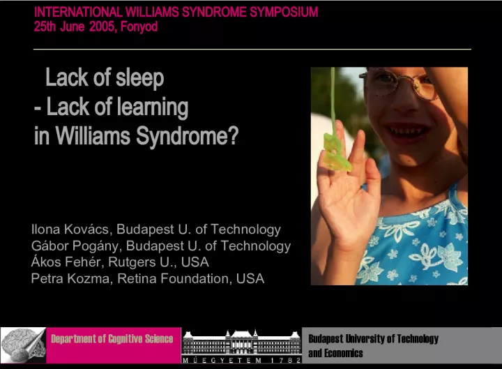
1. Lack of sleep - Lack of learning in Williams Syndrome? INTERNATIONAL WILLIAMS SYNDROME SYMPOSIUM 25th June 200 5 , Fonyod Department of Cognitive Science Budapest University of Technology and Economics Ilona Kovcs, Budapest U. of Technology G bor Pog ny , Budapest U. of Technology kos Fehr, Rutgers U., USA Petra Kozma, Retina Foundation, USA
2. Reiss et al, J Cog Neurosci volume 12 Suppl 1
3. area 17 hi s tometric r esults : s igns of abnormal connectivity Cell measures differ in peripheral visual cortical fields of WS Smaller, more tightly packed cells in most layers on the left side Cell packing density and neuronal size differences may be related to visual spatial deficits in WS Galaburda and Bellugi, J Cog Neurosci vol 12 Suppl 1
4. Primary visual cortex (V1) Local input anal ysi s : L contrast (1 yr) disparity (4 mo) motion (2 mo) color (2 mo) orientation
5. newborn 1mo 2 mo 3 mo 6 mo adult Teller, 1997 Campbell-Robson CSF Chart
6. Local cortical filters in V1 On multiple scales
8. D > 1 D < 1 D = noise spacing / contour spacing
9. (Kovcs and Julesz, PNAS, 1993)
10. Contour integration in 3-month-olds: 60 infants, operant conditioning poor contour integration lack of global integration (closure) (Gerhardstein, Kovcs, Ditre, Fehr Vision Research , 2 00 4)
12. contour integration card test
13. Contour integration in children 510 children , 60 adults Surprisingly slow curve!! 5-6 y 6-7 y 9-10 y 10-11 y 13-14 y 19-30 y D (Kovcs, Kozma, Fehr, Benedek , PNAS , 1999) D = noise spacing / contour spacing
14. contour integration in children: cue specific (no transfer of learning across orientation and color): visual immaturity (not the lack of attention or motivation) dependent on contour spacing: limited cortical connectivity?
15. long axonal connections in V1 , Burkhalter et al , 1993: late maturation of V1 superficial layers horizontal, and V2- V1 feedback connections in humans limited cortical connectivity in children
16. Reiss et al, J Cog Neurosci volume 12 Suppl 1
17. area 17 hi s tometric r esults : s igns of abnormal connectivity Cell measures differ in peripheral visual cortical fields of WMS Smaller, more tightly packed cells in most layers on the left side Cell packing density and neuronal size differences may be related to visual spatial deficits in WMS Galaburda and Bellugi, J Cog Neurosci vol 12 Suppl 1
18. contour integration in Williams Syndrome with C. Pleh, A. Lukacs M. Racsmany Technical U., Budapest P. Kozma Rutgers U., NJ 0.6 0.65 0.7 0.75 0.8 0.85 0.9 6 yr 7 yr 10 yr 11 yr 14 yr 30 yr poor contour integration poor orientation discrimination lack of oblique effect
19. 0 23-24 11-12 shape identification task orientation jitter 5 practice sessions (30 minutes each) visual skill (procedural, habit) learning with P Kozma, and A Feher Rutgers University
20. Groups Mean age Total testing time Sleep 1. (n=8) 24,5 3,5 hours no 2. (n=8) 29,75 10 hours no 3. (n=8) 28,75 5 days yes
22. Day 3. C Day 1. C Day 5. C Day 3. W Day 1. W Day 5. W Day 7. W
23. Texture discrimination task Karni & Sagi 1991: specific to quadrant specific to horizontal background bars specific to eye slow improvement sleep-dependent Scwartz et al, 2002: fMRI; training- dependent monocular increases (V1)
24. Basic skill (implicit, procedural) learning : Time-course of learning (behavioral studies) Plastic changes in the brain (imaging) behaviorally relevant degree of plasticity is retained in the adult mammalian cortex
25. perceptual learning in WS abnormalities in the occipital lobe sleep disorders lack of visual skill learning
27. color defined card
30. lack of closure superiority poor contour integration contour integration in 3-month-old babies
31. Professional musicians - a good model to investigate plastic changes in the human brain Complexity of stimulus Extent of exposure Two steps: - - fast initial phase - - consolidation, and gradual increase in performance Anatomical changes - - Planum temporale - - Anterior corpus callosum - - Primary hand motor and somatosensory - - Cerebellum
32. visual development Things start out badly, then they get better; then, after a long time, they get worse again. (Movshons general law on visual development, Teller & Movshon, 1986)
33. visual development: should follow the maturational pattern of participating cortical structures connectivity supporting low-level spatial integration is immature connectivity supporting the switch between perceptual interpretations is immature is immature top-down connectivity is immature
34. visual development Things start out badly, then they get better; then, after a long time, they get worse again. (Movshons general law on visual development, Teller & Movshon, 1986) Visual development is not a homogeneous process. It might be possible to map it in terms of the maturational pattern of cortical connectivity.
35. 1 mo 2 mo 3-6 mo 7-9 mo > 9 mo (Knaap and Valk, 1990) Stages of myelination
36. ( Thompson et al , 2000) Growth patterns in the developing brain
37. visual development: should follow the maturational pattern of participating cortical structures
38. Patient H.J.A. (Humphreys and Riddoch, 1984, 1987b; Riddoch and Humphreys, 1987a) posterior cerebral artery stroke bilateral lesions of the occipital lobe extending anteriorly towards the temporal lobes dense visual agnosia prosopagnosia alexia without agraphia achromatopsia topographical impairments MRI (1989) : bilateral lesions of inferior temporal gyrus, lateral occipitotemporal gyrus, fusiform gyrus, lingual gyrus (Riddoch et al, Brain, Vol. 122, No. 3, 1999)
39. Eric R. Kandel Nobel in 2000; signal transduction in the nervous system Two steps in synaptic plasticity short-term memory (protein phosphorylation in synapses) long-term memory (protein synthesis, which can lead to alterations in shape and function of the synapse) The switch from short- to long-term memory requires gene expression. (modification of chromatin structure, chromatin is the DNA-protein complex that constitutes chromosomes)
40. Switch from short- to long-term memory in humans Animal models are limited in terms of stimulus complexity and the duration of training. Not clear how mechanisms governing synaptic plasticity at the cellular level are related to the flexibility of operations seen for large-scale neuronal networks. Big questions: Is there plasticity in the adult brain? Are more complex functions relying on the same mechansims of learning?