Signaling pathways in cells

This article explores the various pathways that cells use to communicate, including signaling molecules and receptors, signal transduction pathways, nuclear signaling, cascades, phosphorylation, ATP production, protein responses, and more.
- Uploaded on | 1 Views
-
 folker
About Signaling pathways in cells
PowerPoint presentation about 'Signaling pathways in cells'. This presentation describes the topic on This article explores the various pathways that cells use to communicate, including signaling molecules and receptors, signal transduction pathways, nuclear signaling, cascades, phosphorylation, ATP production, protein responses, and more.. The key topics included in this slideshow are . Download this presentation absolutely free.
Presentation Transcript
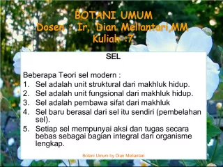
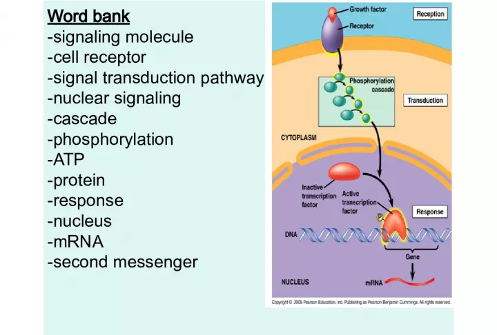
1. Word bank -signaling molecule -cell receptor -signal transduction pathway -nuclear signaling -cascade -phosphorylation -ATP -protein -response -nucleus -mRNA -second messenger
2. Animation • http://bcs.whfreeman.com/thelifewire/conte nt/chp15/15020.html • 1) What is flight or fight? • 2) What is glycogen?
3. 12 days until the final How to use this review: 1) Study notes 2) Do questions without notes 3) For any questions you are stuck on you can look at your notes or phone a friend 4) Use the AP flashcards 5) Make a study group 6) Ask Morris LOTS of questions 7) Know what you know and what you don’t know before the test
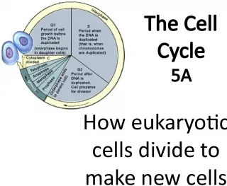
4. THE CELL CYCLE: Chapter 12 Without counting the G 0 phase, a cell cycle takes 12-24 hours for most mammalian cells, and only 20-30 minutes for E. coli cells
5. • http://highered.mcgraw- hill.com/sites/0072495855/student_view0/ch apter2/animation__how_the_cell_cycle_wor ks.html • Take notes on events of each part of the cell cycle • Interphase (G1, S, G2) + PMATC
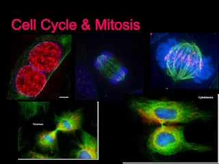
7. Mitosis in the Whitefish blastula Animal mitosis movie
12. Mitosis in Action • Spindle=_________ • Spindle=_________ • Spindle=_________ • Spindle=_________ • Nucleus=_________ • Cell Membrane=______ • Chromosome=______ • Chromosome=______
13. Draw the 9 steps of cell cycle • G1 • S • G2 • Prophase • Prometaphase • Metaphase • Anaphase • Telophase • Cytokinesis
14. Turn to Lab FRQ packet and start question on page 13 -Animal behavior Look at data table a) summarize pattern (2 points) - Identify three physiological or environmental reason that cause this (3 points) • Take out lab report turn in ONLY if you can answer “Yes” to all questions/statements below 1) My discussion is half a page 2) My discussion explain why and not just what happened 3) I used 5 or more voc words
15. The is the 2012 AP Bio Review book. Who wants me to order it for you?
16. I can… • Write about the role of PROTEINS in the cell cycle
17. THE MITOTIC CELL CYCLE The mitotic phase alternates with interphase in the cell cycle Cell Cycle flash animation
18. LE 12-5 G 1 G 2 S (DNA synthesis) INTERPHASE Cytokinesis MITOTIC (M) PHASE Mitosis
19. THE MITOTIC CELL CYCLE The mitotic phase alternates with interphase in the cell cycle Mitosis animation What are the key parts of each phase?
20. The stages of mitotic cell division in an animal cell The light micrographs show dividing lung cells from a newt, which has 22 chromosomes in its somatic cells. The chromosomes appear blue and the microtubules green. (Know the characteristics of the phases)
21. Review the details of each mitotic phase animal cells (Know the characteristics of the phases) Mitosis flash animation (Purves)
22. THE KEY ROLES OF CELL DIVISION • Cell division functions in reproduction, growth, and repair
23. • Cell division distributes identical sets of chromosomes to daughter cells Eukaryotic chromosomes . A tangle of chromosomes (stained orange) is visible within the nucleus of this kangaroo rat epithelial cell.
24. • Every eukaryotic species has a characteristic number of chromosomes in each cell nucleus • Somatic (nonreproductive) cells have two sets of chromosomes • Gametes (reproductive cells: sperm and eggs) have half as many chromosomes as somatic cells • Eukaryotic chromosomes consist of chromatin , a complex of DNA and protein that condenses during cell division
25. • Our DNA is 6 feet long, how does it fit into a nucleus? • Note: 10,000 nuclei fit on the tip of your pencil http://dnalc.org/view/15491-DNA-packaging- 3D-animation-with-narration.html
28. Chromosome duplication and distribution during mitosis . Eukaryotic duplicates each of its multiple chromosomes before it divides. A duplicated chromosome consists of two sister chromatids, which narrow at their centromeres.
29. What do you know about cytoskeleton?
30. The mitotic spindle distributes chromosomes to daughter cells The assembly of spindle microtubules starts in the centrosome, known as a microtubule-organizing center. During interphase, the single centrosome replicates to form two centrosomes. During prophase they form spindle fibers and migrate to the poles.
31. Role of cytoskeleton • http://www.youtube.com/watch?v=5rqbmLiS kpk&feature=related • http://bio.rutgers.edu/~gb101/lab2_mitosis/s ection2_frames.html
34. The mitotic spindle at metaphase • Each of the two joined chromatids of a chromosome has a kinetochore. • Anaphase: proteins holding together the sister chromatids of each chromosome are inactivated and they are now full chromosomes.
35. • Experimental evidence supports the hypothesis that kinetochores use motor proteins that "walk" a chromosome along the attached microtubules toward the nearest pole. • Meanwhile, the microtubules shorten by depolymerizing at their kinetochore ends • In a dividing animal cell, non kinetochore microtubules are responsible for elongating the whole cell during anaphase
36. Cytokinesis divides the cytoplasm How does it differ in animal and plant cells?
37. In animal cells, cytokinesis occurs by cleavage • The cleavage furrow, which begins as a shallow groove in the cell surface. • On the cytoplasmic side, a contractile ring of actin microfilaments and molecules of the protein myosin • The contraction of the dividing cell’s ring of microfilaments is like the pulling of drawstrings Cytokinesis animation
38. Cytokinesis in plant cells has no cleavage furrow During telophase, vesicles derived from the Golgi apparatus move along microtubules to the middle of the cell, where they fuse, producing a cell plate .
39. Mitosis in a plant cell These light micrographs show mitosis in cells of an onion root. How does this differ from animal cell mitosis?
40. Mitosis in eukaryotes may have evolved from binary fission in bacteria Mitosis video (long)
41. A hypothesis for the evolution of mitosis Researchers of eukaryotic cell division have observed in modern organisms what they believe are mechanisms of division intermediate between the binary fission of bacteria and mitosis as it occurs in most eukaryotes.
42. Cancer This man has cancer of the mouth.
43. Regulation of the Cell cycle The timing and rate of cell division in different parts of a plant or animal are crucial to normal growth, development, and maintenance. Do all cells have the same cell cycle? Why is regulation of the cell cycle of interest to research? Cancer Growth Flash animation
44. What is Cancer? • Cancer means uncontrolled cell growth • The body needs to keep cell growth = cell death • Cell cycle checkpoints kill mutated or old cells
45. • http://science.education.nih.gov/supplement s/nih1/cancer/activities/activity2_animations. htm
46. The cell cycle has traffic lights that serve as checkpoints G1 Phase S Phase G2 Phase Mitosis Cytokinesis Is the cell ready for mitosis? Does the body need more cells?
47. Cancer is caused when the checkpoints are broken and the cell cycle keeps going without stopping G1 Phase S Phase G2 Phase Mitosis Cytokinesis
48. What are the types of cancer? *Any part of the body can be cancerous • Skin cancer • Lung cancer • Breast cancer • Testicular cancer • Colon cancer • Liver cancer • Brain cancer Lung Cancer Brain Cancer
50. How do you get cancer? How can you get cancer? • Getting hit in the breast? NO • Having unprotected sex? NO • Smoking? YES • Being in the sun too long? YES
51. Why is cancer so deadly? 1) Mutated cells beat the cell cycle checkpoints and keep dividing 2) They form tumors which then stop your body parts from functioning normally 3) Angiogensis – the tumors hijack blood vessels to keep them alive 4) Metastisis – the cells from the tumor travel and infect other parts of your body *
52. Here is the development of colon cancer.
53. Why is Cancer so Hard to Cure? 1) It is a silent killer, by the time it is found it is already to late 2) Chemo/Radiation therapy can kill cancer cells, but is hard on patients 3) If one cancer cell survives, or travels, cancer will come back
54. Can cancer be prevented? Cancer is not contagious. There is no guaranteed way to prevent cancer, people can reduce their risk (chance) of developing cancer by: A) not using tobacco products B) choosing foods with less fat and eating more vegetables, fruits, and whole grains C) exercising regularly and maintaining a lean weight D) avoiding the harmful rays of the sun, using sunblock, and wearing clothing that protects the skin
55. Mechanical analogy for the cell cycle control system In this diagram of the cell cycle, the flat "stepping stones" around the perimeter represent sequential events. Like the control device of an automatic washer.
56. Cell Cycle Checkpoints • A checkpoint is a critical control point where stop and go-ahead signals can regulate the cycle. • The G1 checkpoint (the "restriction point”) is most important. • If a cell receives a go-ahead signal at the G1 checkpoint, it will usually complete the cycle and divide. • If it does not receive a go-ahead signal at that point, it will exit the cycle, switching into a non-dividing state called the G0 phase. G0 (G zero) resting phase Cell Cycle with Checkpoints Animation
57. Many factors are involved in the regulation of the cell cycle
58. RB inhibits cell division Active Cdk inhibits RB
59. The Cell Cycle Clock: Cyclins and Cyclin-Dependent Kinase • Fluctuations in the abundance and activity of cell cycle control molecules pace the sequential events of the cell cycle. • Protein kinases, give the go-ahead signals at the G1 and G2 checkpoints • The kinases are present at a constant concentration in the growing cell, but much of the time they are in inactive form. • To be active, such a kinase must be attached to a cyclin, a protein that gets its name from its cyclically fluctuating concentration in the cell. • These kinases are called cyclin-dependent kinases, or Cdks. The activity of a Cdk rises and falls with changes in the concentration of its cyclin partner. Cdks are relatively constant Cyclins vary in the cycle
60. Cdks are relatively constant Cyclins vary in the cycle
61. The active enzyme and the activating process can be inhibited by two families of cell cycle inhibitory proteins. 1. Members of the INK4 family bind free CDKs thereby preventing association with cyclins. 2. Members of the CIP family bind and inhibit the active CDK-cyclin complex. http://www.chemsoc.org/exemplarchem/entries/2001/armour/howstrt.htm
62. Internal and external cues help regulate the cell cycle Internal Signals: Messages from the Kinetochores: the APC A gatekeeper at the M phase checkpoint delays anaphase. Regulators from kinetochores insures all the chromosomes are properly attached to the spindle at the metaphase plate and the anaphase-promoting complex (APC) is in an inactive state. When all are attached, the APC then becomes active and indirectly triggers both the breakdown of cyclin and the inactivation of proteins holding the sister chromatids together. Degradation of key regulator proteins such as the anaphase inhibitors PDS1 and CUT2, and the mitosis initiator cyclin B, drives the cell cycle forward.
64. Molecular control of the cell cycle at the G2 checkpoint. The Cdk-cyclin complex called MPF, which acts at the G2 checkpoint to trigger mitosis. The " maturation-promoting factor " triggers the cell’s passage past the G2 checkpoint into M phase Cyclins accumulate during G2 associate with Cdk molecules, the resulting MPF complex initiates mitosis. Later in the M phase, MPF helps switch itself off by initiating a process that leads to the destruction of its cyclin by a protein breakdown mechanism
65. Ubiquitin is part of the pathway for the degradation of proteins
66. Ubiquitin is part of the pathway for the degradation of proteins
67. External Signals: Growth Factors One example of a growth factor is platelet-derived growth factor (PDGF), which is made by blood cells called platelets. The binding of PDGF molecules to these receptors triggers a signal- transduction pathway that leads to stimulation of cell division. The proliferation of fibroblasts helps heal the wounds.
68. Density-dependent inhibition of cell division. Most animal cells also exhibit anchorage dependence Cancer cells exhibit neither density-dependent inhibition nor anchorage dependence
69. Cancer cells have escaped from cell cycle controls Cancer cells do not respond normally to the body’s control mechanisms. They divide excessively and invade other tissues. If unchecked, they can kill the organism. The growth and metastasis of a malignant breast tumor. What is a benign tumor? A malignant tumor? metastasis Breast cancer animation
70. P53 is considered to be a "Guardian of the Genome“ 1. Growth arrest: p21, Gadd45, and 14-3-3 s . 2. DNA repair: p53R2. 3. Apoptosis: Bax, Apaf-1, PUMA and NoxA.
71. P53 re-enforces the G2 checkpoint. This serves as a “tumor suppressor” protein. In the cell, p53 protein binds DNA, which in turn stimulates another gene to produce a protein called p21 that interacts with a cell division-stimulating protein (cdk2). When p21 is complexed with cdk2 the cell cannot pass through to the next stage of cell division. Mutant p53 can no longer bind DNA in an effective way, and as a consequence the p21 protein is not made available to act as the 'stop signal' for cell division. Thus cells divide uncontrollably, and form tumors.
72. http://highered.mcgraw- hill.com/sites/007337797x/student_view 0/chapter9/animation_quiz_- _how_tumor_suppressor_genes_block_ cell_division.html
73. • Explain the following diagram
76. Meiosis
77. Somatic Cells : • body cells • Ex. ___________ • Made by mitosis Gametes : • reproductive cells • Ex. ________
78. Diploid : • Having 2 copies of each chromosome (2n), one from each parent • Somatic cells are diploid • Human diploid number is _____ What are the cells in your body that are diploid? Are gametes diploid? Why or why not? How many chromosomes does a sperm and egg have? Haploid : • Having only 1 copy of each chromosome (n) • Gamete cells are haploid • Human haploid number is _____ What are the cells in your body that are haploid?
79. Copy and fill in the chart below. Organism Diploid # (in somatic cells) Haploid # (in gametes) Cat 19 Rose 12 Goat 30 Rice 24 Dog 39 Chimpanzee 48
81. Eye color gene Homologous pair : • A pair of chromosomes, 1 from mom and 1 from dad • Carry the same genes (ex. eye color gene) • But may contain different information (ex. brown eyes and blue eyes)
82. Mitosis : How our bodies make diploid somatic cells It happens ________________ Meiosis : The special process of making haploid gametes It happens in the ______________ & ______________ Do you do mitosis? Do you do meiosis?
83. Meiosis Video 1 Mitosis vs. Meiosis Video
84. Meiosis
85. Homologous Chromosomes are Homies • They are always the same SIZE • They always have the same type of INFO, but they are not identical
86. Whiteboard Games 1) All members help to find the answer 2) There will be a seat number who will write and a seat number who will present
87. Game 1: Whose my Homie? #1 #2 #3 #4 #5 #6 Seat 2— Writes Seat 3-- Present s
89. Activity • Make 1 set of homologous pairs of chromosomes=2 chromosomes • Put letters on the chromosomes • Demonstrate crossing over • Tips: Use whiteboard and move beads
90. Game 2: Crossing Over • On page 90 all members need to draw crossing over between homologous chromosomes IN COLOR • Book pg 276 Drawing 1—2 homologous chromosomes with letters Drawing 2—Crossing over (twisty style) Drawing 3—Final chromosomes
91. On the bottom of page 90 write • Crossing over occurs between homologous chromosomes • This only occurs in MEIOSIS • Crossing over occurs during prophase 1 and leads to different sperm and egg
92. Dispatch pg 93 • Crossing over is when________________ • Crossing over occurs during____phase of meiosis
93. Mendel’s 2 Laws
94. Independent Assortment • http://www.sumanasinc.com/webcontent/a nimations/content/independentassortment. html
95. On pg 91 write • Mendel’s Law of Independent Assortment — homologous chromosomes line up in different combinations during Metaphase I of Meiosis Draw 2 different alignments
96. Game 3: 2 alignments for these 2 homies E e j J
97. Mendel’s Law 2 pg 92 • Mendel’s Law of Segregation —allele pairs separate during gamete formation and end up in different gametes (sperm and egg) Draw 4 sperm that are segregated
98. Game 4: Segregation or Not? Seat 4— Writes Seat 1-- Present s #1 #2 #3 #4
99. Who won? • Clean up beads, colored pencils, marker and whiteboard • Get ready for exit quiz
100. Exit Quiz 1) Draw a sperm cell that is segregated 2) Draw 2 alignments for homologous chromosomes in metaphase 1
101. Exit Quiz 1) Explain how the cell cycle is regulated 2) How does cancer occur? 3) Give 5 differences between mitosis and meiosis
102. Chapter 12~ The Cell Cycle
103. 2007-2008 Biology is the only subject in which multiplication is the same thing as division …
104. • For reproduction – asexual reproduction • one-celled organisms • For growth – from fertilized egg to multi-celled organism • For repair & renewal – replace cells that die from normal wear & tear or from injury Why do cells divide? amoeba
105. Importance of Cell Division 1. Growth and Development 2. Asexual Reproduction 3. Tissue Renewal Zygote Embryo Fetus Adult 1 Cell 100 cells millions cells 100 trillion cells
106. DNA organization in Prokaryotes • Nucleoid region • Bacterial Chromosome – Single (1) circular DNA – Small • (e.g. E. coli is 4.6X10 6 bp, ~1/100 th human chromosome) • Plasmids – extra chromosomal DNA
107. Bacterial Fission
108. The Cell Cycle • Interphase (90% of cycle) • G1 phase ~ growth • S phase ~ synthesis of DNA • G2 phase ~ preparation for cell division • Mitotic phase • • Mitosis ~ nuclear division • • Cytokinesis ~ cytoplasm division
109. Parts of Cell Cycle • Interphase – G1 – S phase – G2 • M phase – Mitosis (Division of nucleus) • Prophase • Prometaphase • Metaphase • Anaphase • Telophase – Cytokinesis (Division of cytoplasm)
110. Cell Division: Key Roles • Genome : cell’s genetic information • Somatic (body cells) cells • Gametes (reproductive cells): sperm and egg cells • Chromosomes : condensed DNA molecules • Diploid (2n): 2 sets of chromosomes • Haploid (1n): 1 set of chromosomes • Chromatin : DNA-protein complex • Chromatids : replicated strands of a chromosome • Centromere : narrowing “waist” of sister chromatids • Mitosis : nuclear division • Cytokinesis : cytoplasm division • Meiosis : gamete cell division
111. 111 Chromosome Organization • When cells divide, daughter cells must each receive complete copy of DNA • Each cell has about 2 meters of DNA in the nucleus; thin threads called chromatin • Before division, condenses to form chromosomes • DNA also replicates before cell division to produce paired chromatids
112. double- stranded mitotic human chromosomes
113. 113 Normal Karyotype (Fig 18.1)
114. Mitosis • Prophase • Prometaphase • Metaphase • Anaphase • Telophase
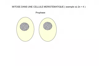
115. Prophase • Chromatin condenses – visible chromosomes • chromatids • Centrioles move to opposite poles of cell – animal cell • Protein fibers cross cell to form mitotic spindle – microtubules • Nucleolus disappears • Nuclear membrane breaks down
116. Prometaphase – spindle fibers attach to centromeres • creating kinetochores – microtubules attach at kinetochores • connect centromeres to centrioles – chromosomes begin moving
117. Metaphase • Centrosomes at opposite poles • Centromeres are aligned • Kinetochores of sister chromatids attached to microtubules (spindle)
119. Anaphase • Paired centromeres separate; sister chromatids liberated • Chromosomes move to opposite poles • Each pole now has a complete set of chromosomes
120. Separation of chromatids • In anaphase, proteins holding together sister chromatids are inactivated – separate to become individual chromosomes 2 chromosomes 1 chromosome 2 chromatids single-stranded double-stranded
121. • Kinetochores use motor proteins that “walk” chromosome along attached microtubule – microtubule shortens by dismantling at kinetochore (chromosome) end Chromosome movement
122. Telophase • Daughter nuclei form • Nuclear envelopes arise • Chromatin becomes less coiled • Two new nuclei complete mitosis • Cytokinesis begins – cell division
125. Mitosis in whitefish blastula
127. Cytokinesis • Cytoplasmic division • Animals – constriction belt of actin microfilaments around equator of cell • cleavage furrow forms • splits cell in two • like tightening a draw string
128. Cytokinesis in Plants • Plants – cell plate forms • vesicles line up at equator – derived from Golgi • vesicles fuse to form 2 cell membranes – new cell wall laid down between membranes • new cell wall fuses with existing cell wall
129. onion root tip
130. Any Questions??
131. Cell Cycle regulation • Checkpoints – cell cycle controlled by STOP & GO chemical signals at critical points – signals indicate if key cellular processes have been completed correctly
132. Checkpoint control system • 3 major checkpoints: – G 1 /S • can DNA synthesis begin? – G 2 /M • has DNA synthesis been completed correctly? • commitment to mitosis – spindle checkpoint • are all chromosomes attached to spindle? • can sister chromatids separate correctly?
133. G 1 /S checkpoint • G 1 /S checkpoint is most critical – primary decision point • “ restriction point ” – if cell receives “GO” signal , it divides • internal signals: cell growth (size), cell nutrition • external signals: “growth factors” – if cell does not receive signal, it exits cycle & switches to G 0 phase • non-dividing, working state
134. “Go-ahead” signals • Protein signals that promote cell growth & division – internal signals • “ promoting factors ” – external signals • “ growth factors ” • Primary mechanism of control – phosphorylation • kinase enzymes • either activates or inactivates cell signals
135. Cell cycle signals • Cell cycle controls – cyclins • regulatory proteins • levels cycle in the cell – Cdks • cyclin-dependent kinases • phosphorylates cellular proteins – activates or inactivates proteins – Cdk-cyclin complex • triggers passage through different stages of cell cycle activated Cdk inactivated Cdk
136. External signals • Growth factors – coordination between cells – protein signals released by body cells that stimulate other cells to divide • density-dependent inhibition – crowded cells stop dividing – each cell binds a bit of growth factor » not enough activator left to trigger division in any one cell • anchorage dependence – to divide cells must be attached to a substrate » “touch sensor” receptors
137. Growth Factors and Cancer • Growth factors can create cancers – proto-oncogenes • normally activates cell division – growth factor genes – become oncogenes (cancer-causing) when mutated • if switched “ON” can cause cancer • example: RAS (activates cyclins) – tumor-suppressor genes • normally inhibits cell division • if switched “OFF” can cause cancer • example: p53
138. Cancer & Cell Growth • Cancer is essentially a failure of cell division control – unrestrained, uncontrolled cell growth • What control is lost? – lose checkpoint stops – gene p53 plays a key role in G 1 /S restriction point • p53 protein halts cell division if it detects damaged DNA – options: » stimulates repair enzymes to fix DNA » forces cell into G 0 resting stage » keeps cell in G 1 arrest » causes apoptosis of damaged cell • ALL cancers have to shut down p53 activity p53 discovered at Stony Brook by Dr. Arnold Levine p53 is the Cell Cycle Enforcer
139. DNA damage is caused by heat, radiation, or chemicals. p53 allows cells with repaired DNA to divide. Step 1 DNA damage is caused by heat, radiation, or chemicals. Step 1 Step 2 Damaged cells continue to divide. If other damage accumulates, the cell can turn cancerous. Step 3 p53 triggers the destruction of cells damaged beyond repair. ABNORMAL p53 NORMAL p53 abnormal p53 protein cancer cell Step 3 The p53 protein fails to stop cell division and repair DNA. Cell divides without repair to damaged DNA. Cell division stops, and p53 triggers enzymes to repair damaged region. Step 2 DNA repair enzyme p53 protein p53 protein p53 — master regulator gene
140. Development of Cancer • Cancer develops only after a cell experiences ~6 key mutations (“hits”) – unlimited growth • turn on growth promoter genes – ignore checkpoints • turn off tumor suppressor genes (p53) – escape apoptosis • turn off suicide genes – immortality = unlimited divisions • turn on chromosome maintenance genes – promotes blood vessel growth • turn on blood vessel growth genes – overcome anchor & density dependence • turn off touch-sensor gene It ’ s like an out-of-control car with many systems failing !
141. What causes these “hits”? • Mutations in cells can be triggered by UV radiation chemical exposure radiation exposure heat cigarette smoke pollution age genetics
142. Tumors • Mass of abnormal cells – Benign tumor • abnormal cells remain at original site as a lump – p53 has halted cell divisions • most do not cause serious problems & can be removed by surgery – Malignant tumor • cells leave original site – lose attachment to nearby cells – carried by blood & lymph system to other tissues – start more tumors = metastasis • impair functions of organs throughout body
143. Cancer: breast cancer cell & mammogram
144. Traditional treatments for cancers • Treatments target rapidly dividing cells – high-energy radiation • kills rapidly dividing cells – chemotherapy • stop DNA replication • stop mitosis & cytokinesis • stop blood vessel growth
145. New “miracle drugs” • Drugs targeting proteins (enzymes) found only in cancer cells – Gleevec • treatment for adult leukemia (CML) & stomach cancer (GIST) • 1st successful drug targeting only cancer cells Novartes without Gleevec with Gleevec
146. 2008-2009 Any Questions??