Body Membranes: Skin and Mucous Membranes

This chapter focuses on body membranes that cover surfaces, line body cavities, and form protective sheets. There are two groups of body membranes, classified according to tissue
- Uploaded on | 1 Views
-
 jeremybrewer
About Body Membranes: Skin and Mucous Membranes
PowerPoint presentation about 'Body Membranes: Skin and Mucous Membranes'. This presentation describes the topic on This chapter focuses on body membranes that cover surfaces, line body cavities, and form protective sheets. There are two groups of body membranes, classified according to tissue. The key topics included in this slideshow are . Download this presentation absolutely free.
Presentation Transcript
Slide1Chapter 4Skin and Body Membranes
Slide2Body Membranes• Cover surfaces, line body cavities, form protective sheets • 2 groups – classified according to tissue
Slide3Epithelial membranes• Aka covering and lining membranes • Do contain some connective tissue • Considered simple organs • (1) Cutaneous membrane – Skin – Dry membrane
Slide4•(2) Mucous membranes (mucosa) – Epithelium on top of lamina propria – Lines any cavity w/ an exterior opening – Wet (moist) membranes – Continuously covered by secretions – Ex. Respiratory, digestive, urinary, reproductive tracts – Adapted for absorption or secretion
Slide5•(3) Serous membranes (serosa) – Simple squamous epithelium on areolar tissue – Line cavities closed to exterior – Occur in pairs • Parietal layer – wall of ventral body cavity • Visceral layer – covers organs in the cavity
Slide6–Serous fluid – btw layers – secreted by both layers – Allows organs to move w/out friction (heart,stomach) – Name depends on location • Abdominal cavity – peritoneum; lungs – pleura; heart - pericardium
Slide7Connective membranes• Aka synovial membranes • Areolar tissue – no epithelial • Line fibrous capsules around joints – Smooth surface – Secrete lubricating fluid
Slide8•Line bursae (small sacs of connective tissue) and tendon sheaths • Cushion organs that move against each other
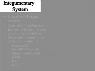
Slide9Integumentary System Cutaneous membrane • Aka integument – “covering” • 2 layers – epidermis & dermis – usually very close – Burns/friction may cause separation = blister • Functions – Insulates and cushions deeper organs – Regulates body temp – capillaries, sweat glands – Mini excretory system – urea, uric acid, salts, water released w/ sweat – Synthesizes immunity proteins – Synthesizes vitamin D – Contains cutaneous receptors – touch, pressure, temp, pain
Slide10Epidermis• Avascular • Keratinocytes – majority of cells – produce keratin (tough, protective, waterproofing) • May have up to 5 strata (layers)
Slide11•(1) Stratum basale – Deepest – Best nourished via diffusion – Aka stratum germinativum because they are continually dividing – Daughter cells are pushed upward – Contains melanocytes – produce melanin (pigment) – Sunlight stimulates production – Concentrated in 1 spot = freckles, moles
Slide12•(2) Stratum spinosum • (3) Stratum granulosum • (4) Stratum lucidum – Dead – unable to get nutrients and oxygen – Occurs in hairless, extra thick areas – palms, soles of feet • (5) Stratum corneum – 20-30 cell layers (3/4 of epidermis) – Dead – completely filled w/ keratin – Aka cornified cells (corno = horn) – Rubbed/flakes off and is replaced by lower cells – Cycle 25-45 days
Slide14Dermis• Hide – leathergoods • Strong, stretchy envelope that holds body together • 2 layers • (1) Papillary layer – Upper dermal – Dermal papillae – projections on superior surface – Contain capillary loops which provide nutrients – May have Meissner’s corpuscles – touch receptors – Form ridges - fingerprints
Slide15•(2) Reticular layer – Deepest skin layer – Contains blood vessels, sweat/oil glands, Pacinian corpuscles (pressure receptors) • Both layers contain phagocytes that work to prevent bacteria from going deeper • If blood supply is restricted = cell death = skin ulcers (bed sores) – Ex. Decubitus ulcers – found in bedridden patients
Slide16•Collagen fibers – provide toughness – Attract/bind water – keep skin hydrated • Elastic fibers – give skin elasticity – # of fibers decreases – allows wrinkles • Blood vessels help maintain temp – To release heat – vessels swell – To conserve heat – vessels constrict, may bypass capillaries
Slide17Hypodermis• Subcutaneous tissue – adipose tissue • Not part of skin • Acts as an anchor, shock absorber, insulator
Slide18Skin Color• 3 pigments – Melanin – yellow, brown, black – Carotene – yellow-orange – Oxygen-rich hemoglobin – red/pink • Emotional stimuli or disease affect color • Redness (erythema) – Blushing, fever, inflammation, allergy
Slide19•Pallor (blanching) – become pale – Anemia, low blood pressure, fear • Jaundice (yellow cast) – Liver disorder – bile deposited in tissues
Slide20•Bruises (hematomas) – Where blood has left vessels and clotted in tissue space – May be vitamin C deficit or hemophilia
Slide21•Cyanosis – Bluish cast due to low oxygen – Common in people with breathing disorders