UCLA Head & Neck Surgery Resident Lecture Series: A Little History with Dr. Marc Cohen
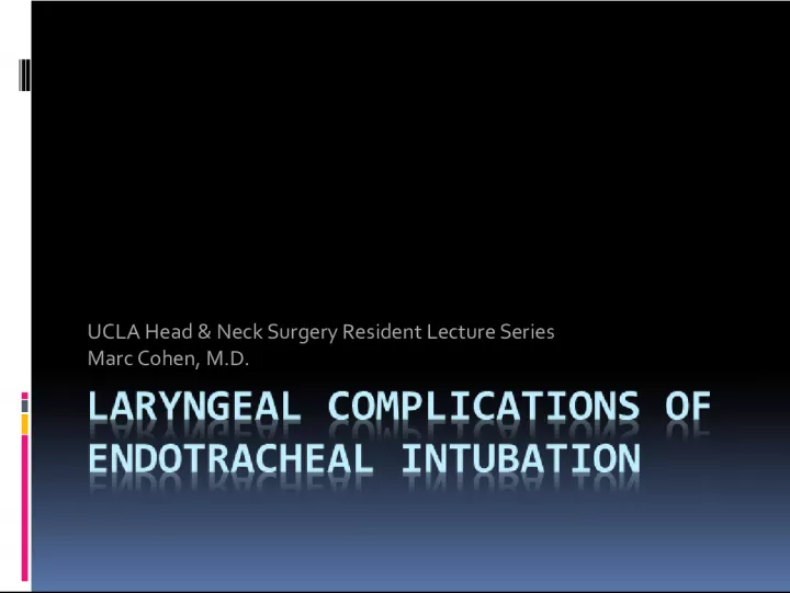

In this video tutorial, Dr. Marc Cohen of UCLA's Head & Neck Surgery program delves into a little bit of history behind the field, offering valuable insights to current residents.
- Uploaded on | 2 Views
-
 olgaieveckiy
About UCLA Head & Neck Surgery Resident Lecture Series: A Little History with Dr. Marc Cohen
PowerPoint presentation about 'UCLA Head & Neck Surgery Resident Lecture Series: A Little History with Dr. Marc Cohen'. This presentation describes the topic on In this video tutorial, Dr. Marc Cohen of UCLA's Head & Neck Surgery program delves into a little bit of history behind the field, offering valuable insights to current residents.. The key topics included in this slideshow are . Download this presentation absolutely free.
Presentation Transcript
1. UCLA Head & Neck Surgery Resident Lecture Series Marc Cohen, M.D.
2. A little history
3. Video tutorial
4. A little history
5. A little history By 1910, intubation for anesthesia had become an accepted practice During WWI, Magill and Macintosh made profound improvements In 1970, high-volume, low pressure cuffs were introduced
6. Prolonged intubation vs. tracheotomy? In the 1960s, long term intubation for the management of premature LBW infants was recommended Until. Subglottic stenosis was recognized
7. Indications for endotracheal intubation 1. Temporary relief of upper airway obstruction 2. Assisted ventilation for respiratory failure 3. Pulmonary toilet
8. What are the potential complications of endotracheal intubation? Edema Granuloma Healed fibrous nodule Interarytenoid adhesion Posterior glottic stenosis Subglottic stenosis Healed furrows Ductal cysts Hematoma Laceration Subluxation of arytenoid cartilage Loss of mobility of cricoarytenoid joint Vocal cord paralysis Nasogastric tube syndrome
9. Pathogenesis Pressure-Induced Injuries Vulnerable structures Medial surfaces of arytenoids Vocal processes Cricoarytenoid joints Cricoid cartilage Posterior glottic/Interarytenoid region
10. Pathogenesis Supraglottic structures may become edematous, but rarely sustain serious damage Tracheal injuries have also become less significant due to low pressure cuffs Although there is potential for injury if the cuff is inflated too high
11. Pathogenesis The microcirculation of the mucosa and mucoperichondrium is interrupted when pressure from the ETT exceeds capillary pressure Ischemia Necrosis Edema, Hyperemia, Ulceration, and Erosion
12. Factors for susceptibility Extrinsic factors Diameter of ETT Duration of intubation Traumatic or multiple intubations Patient factors Poor tissue perfusion (i.e. sepsis, organ failure, etc) LPR Abnormal larynx Wound healing, keloid Movement During ventilator use During suctioning During coughing During transport
13. Laryngeal Bedsore Superficial ulceration can occur within hours of intubation Usually heals without scarring As ETT pressure continues, migration of inflammatory cells ensues If epithelial erosions are incomplete, epithelium may be replaced by squamous metaplasia Further pressure causes ulceration through mucosa to cartilage Causes perichondritis and destructive chondritis As opposed to superficial damage, deeper ulceration heals by secondary intention and fibrosis
14. Edema 3 locations 1. Reinkes space Usually persists after extubation 2. Ventricular mucosa, seen as protrusion Usually resolves after extubation 3. Subglottis Usually resolves after extubation
15. Edema
16. Granulation tissue Seen within 48 hours Proliferate at periphery of ulcerated areas
17. Pathogenesis
18. Granulation tissue Flaps of granulation tissue Can move with inspiration/expiration Inspiratory stridor Not recommended to excise both sides Most cases will resolve without any intervention once ETT is removed
19. Granulation tissue Incomplete resolution of granulation tissue can yield: Postintubation granuloma Healed fibrous nodule
20. Interarytenoid adhesion
21. Posterior glottic stenosis Forms when scar contracts after wide ulceration with no intact median strip of mucosa Vocal cords unable to abduct Glottis remains partly closed Inspiratory stridor Voice is usually unaffected Treatment: deep vertical division with laser or 11 blade down to level of cricoid Re-stenosis is likely Costal cartilage graft may be necessary (endoscopically or open)
22. Posterior glottic stenosis
23. Subglottic stenosis Many causes In infants, most common factors related to acquired SS are ETT size and LPR during long-term intubation Presentation in an infant: Failed extubation Recurrent or atypical croup Slowly progressive airway obstruction Difficulty passing ETT Postanesthesia stridor
24. Cotton-Myer Grading System Grade I - < 50 % obstruction Grade II 51-70% obstruction Grade III 71-99% obstruction Grade IV No detectable lumen Rule of thumb: Subglottic diameter < 4.0 mm in a full-term infant is the lower limit of normal (< 3.0 mm in a preterm infant)
25. Subglottic stenosis When repeated attempts at extubation fail: Reintubate with smaller ETT Racemic epinepherine Dexamethasone If these maneuvers fail: Cricoid split with/without cartilage graft Tracheostomy
26. Ductal Cysts Result from retention of mucus in obstructed, dilated ducts of submucosal mucous glands Most are small and require no treatment When large and cause obstruction, endoscopic removal is required
27. Ductal cysts
28. Arytenoid dislocation May occur during passage of an ETT Left arytenoid is usually affected since intubation occurs from right side of mouth Patient will complain of hoarseness, throat discomfort, odynophagia, and cough Microlaryngoscopy and closed reduction should be performed early
29. Arytenoid dislocation
30. Nasogastric tube syndrome Occurs when NGT rests centrally, rather than laterally Anterior wall of hypopharynx/posterior wall of cricoid becomes ulcerated Results in perichondritis, chondritis, necrosis Can progress to sudden, life- threatening bilateral vocal cord paralysis due to myositis of PCA muscles Diabetics and renal transplants who are in renal failure are especially vulnerable Warning signs: hoarseness, otalgia, and odynophagia Treatment: remove NGT, abx, G-tube, and possible tracheostomy
31. Timeline of postextubation obstruction Immediate: flaps of granulation tissue, laryngeal spasm Minutes to hours: flaps of granulation tissue, subglottic edema, granulation tissue, LPR Days to weeks: persistent edema or granulation tissue, granuloma Months: posterior glottic stenosis, subglottic stenosis
32. To trach or not to trach? One school of thought is that anyone who is intubated longer than 7 days should undergo tracheotomy Newer recommendations are for DL after 7 days if no evidence of significant laryngeal pathology, keep the patient intubated unless plan for long-term tracheostomy