Introduction to Serology

Learn to determine if a stain is blood and whether it is of human or animal origin through this course on Serology.
- Uploaded on | 4 Views
-
 olivia
About Introduction to Serology
PowerPoint presentation about 'Introduction to Serology'. This presentation describes the topic on Learn to determine if a stain is blood and whether it is of human or animal origin through this course on Serology.. The key topics included in this slideshow are . Download this presentation absolutely free.
Presentation Transcript
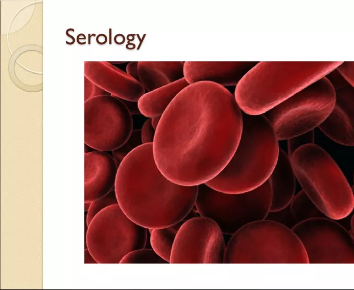
1. Serology Serology
2. Objectives Objectives You will be able to: Determine whether a stain is blood. Determine whether a bloodstain is human or animal blood. Determine the blood type of a simulated bloodstain using the ABO/Rh system. Explore bloodstain patterns as a function of velocity, direction, and height of fall. Use technology and mathematics to improve investigations and communications.
3. Serology Serology The examination and analysis of body fluids. A forensic serologist may analyze a variety of body fluids including saliva, semen, urine, and blood. From 1950 to the late 1980s, forensic serology was a most important part of lab procedures. With DNA techniques, more time, money, and significance were placed on developing DNA labs. With limited funds and the time required for DNA testing, most labs still use many of the basic serology testing procedures
4. What makes up our blood? What makes up our blood? RED BLOOD CELLS (Erythrocytes) The most abundant cells in our blood; they are produced in the bone marrow and contain a protein called hemoglobin that carries oxygen to our cells. WHITE BLOOD CELLS (Leukocytes ) They are part of the immune system and destroy infectious agents called pathogens. PLASMA This is the yellowish liquid portion of blood that contains electrolytes, nutrients and vitamins, hormones, clotting factors, and proteins such as antibodies to fight infection. PLATELETS (Thrombocytes ) The clotting factors that are carried in the plasma; they clot together in a process called coagulation to seal a wound and prevent a loss of blood.
5. Blood Characteristics Blood Characteristics Plasma is the fluid portion of the blood (55 percent). Cells (45 percent) Erythrocytes are red blood cells. They are responsible for oxygen distribution. Leukocytes are the white blood cells; they are responsible for cleaning the system of foreign invaders. Thrombocytes or platelets are responsible for blood clotting. Serum is the liquid that separates from the blood when a clot is formed
6. Blood Terminology Blood Terminology ABO blood groups based on having A, B, both, or no antigens on red blood cells Rh factor may be present on red blood cells; positive if present and negative if not Antigen a substance that can stimulate the body to make antibodies. Certain antigens (proteins) found in the plasma of the red blood cells membrane account for blood type. Antibody a substance that reacts with an antigen Agglutination clumping of red blood cells; will result if blood types with different antigens are mixed
7. Forensic Science Lab Activity T. Trimpe 2006 http://sciencespot.net/ Warning: Some material in this presentation and related videos may be too graphic for some people.
8. Unknown Stain at a Scene Unknown Stain at a Scene Questions to be answered: Is it blood? Is it human blood? Whose is it? Determine blood type, alcohol content, drugs present Determine the method(s) in which blood may have been deposited
9. Presumptive Tests for Blood Determination Presumptive Tests for Blood Determination Kastle-Meyer color test a mixture of phenolphthalein and hydrogen peroxide; the hemoglobin will cause the formation of a deep pink color if blood is present Hematest tablet reacts with the heme group in blood, causing a blue-green color Luminol test reaction with blood to produce light
10. Human versus Animal Blood Human versus Animal Blood Microscopic observation Precipitin test blood is injected into a rabbit; antibodies are formed; the rabbits blood is extracted as an antiserum; the antiserum is placed on sample blood. The sample will react with human proteins if human blood is present. This test is very sensitive and requires only a small amount of blood.
11. Animal Blood Animal Blood Larger nucleic red blood cells Frog blood
12. Microscopic Views Microscopic Views Bird Blood Cat Blood Dog Blood Fish Blood Frog Blood Snake Blood Human Blood Horse Blood
14. Blood Spatter Evidence Blood Spatter Evidence A field of forensic investigation that deals with: physical properties of blood patterns produced under different conditions as a result of various forces being applied to the blood. Blood, as a fluid, follows the laws of physics.
15. Blood Spatter Evidence Blood Spatter Evidence Blood samples Can be analyzed to determine blood type and DNA, which can be matched to possible suspects. Blood droplets Can be analyzed to give clues to the location of a crime, movement of a victim, and type of weapon. Blood spatter Can be analyzed to determine patterns that give investigators clues to how a crime might have happened.
16. Light Source Investigators will first examine the crime scene to look for areas that may contain blood. They may use a high-intensity light or UV lights to help them find traces of blood as well as other bodily fluids that are not visible under normal lighting conditions. How is blood evidence detected at a crime scene? Blood Reagent Tests These tests, referred to as presumptive tests , are used to detect blood at crime scenes based upon the properties of hemoglobin in the blood. Further tests at the crime lab can determine if it is human blood or not. Examples : Phenolphthalein is a chemical that is still utilized today and is usually referred to as the Kastle-Meyer test and produces a pink color when it reacts with hemoglobin. HemaStix is a strip that has been coated with tetramethylbenzidine (TMB) and will produce a green or blue- green color with the presence of hemoglobin. Kastle-Meyer Test Video HemaStix
17. Luminol This chemical is used by crime scene investigators to locate traces of blood, even if it has been cleaned or removed. Investigators spray a luminol solution is throughout the area under investigation and look for reactions with the iron present in blood, which causes a blue luminescence . One problem is that other substances also react, such as some metals, paints, cleaning products, and plant materials. Another problem is that the chemical reaction can destroy other evidence in the crime scene. Luminol Reaction LCV or Leuco Crystal Violet , is one type of chemical process that is used for blood enhancement. Using this test helps to make the blood evidence more visible so it can be photographed and analyzed. Fluorescein This chemical is also capable of detecting latent or old blood, similar to luminol. It is ideal for fine stains or smears found throughout a crime scene. After the solution has been sprayed onto the substance or area suspected to contain blood, a UV light and goggles are used to detect any illuminated areas, which appear greenish-white if blood is present. It may also react to many of the same things as luminol (copper and bleach). Fluorescein Reaction in UV Light
18. What does the abbreviation BPA represent? B loodstain P attern A nalysis What can an investigator learn from the analysis of a blood spatter? Type and velocity of weapon Number of blows Handedness of assailant (right or left-handed) Position and movements of the victim and assailant during and after the attack Which wounds were inflicted first Type of injuries How long ago the crime was committed Whether death was immediate or delayed http://www.crimescenetwo.com/img/popup/book2p2.jpg Source: http://science.howstuffworks.com/bloodstain-pattern-analysis1.htm
19. Bloodstain Pattern Analysis Terms Bloodstain Pattern Analysis Terms Spatter Bloodstains created from the application of force to the area where the blood originated. Origin/Source The place from where the blood spatter came from or originated. Angle of Impact The angle at which a blood droplet strikes a surface. Parent Drop Spines Satellite Spatters Parent Drop The droplet from which a satellite spatter originates. Satellite Spatters Small drops of blood that break of from the parent spatter when the blood droplet hits a surface. Spines The pointed edges of a stain that radiate out from the spatter; can help determine the direction from which the blood traveled.
20. Types of Bloodstain Patterns Types of Bloodstain Patterns Passive Bloodstains Patterns created from the force of gravity Drop, series of drops, flow patterns, blood pools , etc. Projected Bloodstains Patterns that occur when a force is applied to the source of the blood Includes low, medium, or high impact spatters, cast- off, arterial spurting, expiratory blood blown out of the nose, mouth, or wound. Transfer or Contact Bloodstains These patterns are created when a wet, bloody object comes in contact with a target surface; may be used to identify an object or body part. A wipe pattern is created from an object moving through a bloodstain, while a swipe pattern is created from an object leaving a bloodstain. Images from http://www.bloodspatter.com/BPATutorial.htm
21. Blood Pattern Reconstruction Blood Pattern Reconstruction Scene Pattern Reconstruction 1. Stain condition 2. Pattern 3. Distribution 4. Location 5. Directionality Lab Results Reconstruction 1. Genetic marker typing 2. Age determination 3. Source determination 4. Race determination 5. Sex determination