Understanding the Urinary System: Balancing Water, Ions, and pH

The urinary system is a vital component of the human body that is responsible for maintaining the balance of water and ions in the blood, as well as regulating
- Uploaded on | 15 Views
-
 polyan
About Understanding the Urinary System: Balancing Water, Ions, and pH
PowerPoint presentation about 'Understanding the Urinary System: Balancing Water, Ions, and pH'. This presentation describes the topic on The urinary system is a vital component of the human body that is responsible for maintaining the balance of water and ions in the blood, as well as regulating. The key topics included in this slideshow are . Download this presentation absolutely free.
Presentation Transcript
Slide1The Urinary System The Urinary System
Slide2OverviewOverview The urinary system balances the water content in the blood as well as ions of Na and K and pH. The urinary system balances the water content in the blood as well as ions of Na and K and pH. It Regulates the volume and composition of the fluids and excrete unwanted materials. It Regulates the volume and composition of the fluids and excrete unwanted materials.
Slide3Anatomy of the Urinary System Anatomy of the Urinary System
Slide4Internal Kidney Structure Internal Kidney Structure
Slide5Urinary Bladder Anatomy Urinary Bladder Anatomy Trigone Rugae- Flods of the bladder Detrusor Muscle – smooth muscle Urethra Urethral Sphincter -Involuntary
Slide6Physiology of the bladder function Physiology of the bladder function The bladder stretches as it fills with urine and when there is 250mL you feel the need to void. The bladder stretches as it fills with urine and when there is 250mL you feel the need to void. Voiding begins with voluntary relaxation of the external urethral sphincter muscle. Voiding begins with voluntary relaxation of the external urethral sphincter muscle. Once you relax the urethral sphincter the involuntary response of contraction takes over. AKA micturition Once you relax the urethral sphincter the involuntary response of contraction takes over. AKA micturition Inability to void because kidneys are not excreting urine is known as suppression . Inability to void because kidneys are not excreting urine is known as suppression . Inability to void even though urine is present in the bladder is known as retention. Inability to void even though urine is present in the bladder is known as retention. Involuntary micturition = Incontinance Involuntary micturition = Incontinance
Slide7 Urethra Urethra Small tube lined with mucous membrane leading from the floor of the bladder to the exterior of the body. Small tube lined with mucous membrane leading from the floor of the bladder to the exterior of the body. In females this distance is about 3cm. In females this distance is about 3cm. In males this distance extends along a winding path for about 20 cm and passes through the middle of the prostate gland just after leaving the bladder. & is shared as part of the reproductive system. In males this distance extends along a winding path for about 20 cm and passes through the middle of the prostate gland just after leaving the bladder. & is shared as part of the reproductive system.
Slide8Microscopic Structure of the functional unit of a kidney is the Nephron. Microscopic Structure of the functional unit of a kidney is the Nephron. Bowman’s Capsule Proximal convoluted tubule Loop of Henle Distal convoluted tubule Collecting duct There are approximately 1.25 million nephrons Per Kidney. Components of the Nephron
Slide9Glomeruli – Many glomerulus Glomeruli – Many glomerulus Glomerular capillaries
Slide10Bowman’s Capsule + Glomerulus = Renal corpuscle Bowman’s Capsule + Glomerulus = Renal corpuscle
Slide11The loop of Henle dips into the medulla into a renal pyramid. The loop of Henle dips into the medulla into a renal pyramid.
Slide12The collecting ducts empty into the calyces which empty into the renal pelvis. The collecting ducts empty into the calyces which empty into the renal pelvis.
Slide13Blood vessels of the Kidneys Blood vessels of the Kidneys About 1200 ml of blood flows through the kidneys every minute About 1200 ml of blood flows through the kidneys every minute Which means approximately 1/5 of all the blood pumped by the heart per minute goes to the kidneys. Which means approximately 1/5 of all the blood pumped by the heart per minute goes to the kidneys. Usual direction of blood flow is Usual direction of blood flow is Arteries-Arterioles-Capillaries-venules-veins Arteries-Arterioles-Capillaries-venules-veins
Slide14Blood flow through Kidney tissue follows this path… Blood flow through Kidney tissue follows this path… Renal Artery Renal Artery Interlobular artery-penetrate the cortex Interlobular artery-penetrate the cortex Afferent arteriole Afferent arteriole Glomerulus Glomerulus Efferent arteriole Efferent arteriole Peritubular capillaries(vasa recta) Peritubular capillaries(vasa recta) Venules Venules Interlobular vein Renal Vein Interlobular vein Renal Vein
Slide15Function of the kidney Function of the kidney KIDNEYS… KIDNEYS… Process blood plasma and excrete urine. Process blood plasma and excrete urine. Most important organs in the body for maintaining fluid-electrolyte and acid-base balances. Most important organs in the body for maintaining fluid-electrolyte and acid-base balances. Excrete Nitrogenous wastes from protein metabolism. (urea) Excrete Nitrogenous wastes from protein metabolism. (urea)
Slide16Kidney Failure means homeostatic failure and Kidney Failure means homeostatic failure and If not relieved, If not relieved, inevitable DEATH. inevitable DEATH.
Slide17Kidneys also… Kidneys also… Influence the rate of secretion of the hormones Influence the rate of secretion of the hormones ADH – Antidiuretic hormone ADH – Antidiuretic hormone Aldosterone Aldosterone and synthesize erythropoietin –active form of vitamin D and prostaglandins. and synthesize erythropoietin –active form of vitamin D and prostaglandins.
Slide18Formation of urine Formation of urine Urine is formed by 3 means Urine is formed by 3 means 1. Filtration – the movement of water and solutes from the plasma in the glomerulus, across the glomerular-capsular membrane, and into the capsular space of the Bowman’s capsule 1. Filtration – the movement of water and solutes from the plasma in the glomerulus, across the glomerular-capsular membrane, and into the capsular space of the Bowman’s capsule
Slide192. Reabsorption - movement of molecules out of the tubule and into the peritubular blood. 2. Reabsorption - movement of molecules out of the tubule and into the peritubular blood.
Slide203. Secretion – movement of the molecules out of the peritubular blood into the tubule for excretion. 3. Secretion – movement of the molecules out of the peritubular blood into the tubule for excretion.
Slide21FiltrationFiltration Occurs through the glomerular capiliaries to the Bowman’s Capsule Occurs through the glomerular capiliaries to the Bowman’s Capsule Large substances are separated from small substances. Large substances are separated from small substances. Blood cells and blood proteins are typically TOO large to filter through the glomerulus so as the blood leaves the glomerulus in the efferent arteriole it is relatively clean and has lost most of its fluid. Blood cells and blood proteins are typically TOO large to filter through the glomerulus so as the blood leaves the glomerulus in the efferent arteriole it is relatively clean and has lost most of its fluid.
Slide22Blood cells are too large to filter out. So what does filter out? Blood cells are too large to filter out. So what does filter out? Water Water Salts Salts Bicarbonate Bicarbonate H+ H+ Urea Urea Glucose Glucose Amino acids Amino acids Some drugs Some drugs
Slide23Returned to the blood immediately: Reabsorption occurs mostly in the PCT. Returned to the blood immediately: Reabsorption occurs mostly in the PCT. Water and ions Water and ions Glucose and amino acids Glucose and amino acids Passive and Active transport mechanisms are at work in the tubules. Passive and Active transport mechanisms are at work in the tubules. MANY Carriers are available for materials that need to be reclaimed. MANY Carriers are available for materials that need to be reclaimed. Very few carriers for things that are of little or no use to the body. Very few carriers for things that are of little or no use to the body.
Slide24Reabsorption in Reverse: Secretion Reabsorption in Reverse: Secretion And it gets rid of substances not already in the filtrate such as drugs. And it gets rid of substances not already in the filtrate such as drugs. H+ and K+ and creatinine move from the peritubular blood to the tubule. H+ and K+ and creatinine move from the peritubular blood to the tubule.
Slide25Urine vs. Filtrate Urine vs. Filtrate Filtrate contains everything that blood plasma does (except proteins) Filtrate contains everything that blood plasma does (except proteins) Most of the Water, nutrients and necessary ions are reabsorbed by the time it reaches the collecting ducts. Most of the Water, nutrients and necessary ions are reabsorbed by the time it reaches the collecting ducts. Urine contains most of the waste and unneeded substances. Urine contains most of the waste and unneeded substances.
Slide26In 24 hours In 24 hours 150 to 180 liters of blood plasma filter through the kidneys (nephrons) 150 to 180 liters of blood plasma filter through the kidneys (nephrons) Only 1 to 1.8 liters of urine are produced. Only 1 to 1.8 liters of urine are produced. WHAT HAPPENS TO MOST OF THE FILTRATE? WHAT HAPPENS TO MOST OF THE FILTRATE? Initial Filtrate –dilute Initial Filtrate –dilute Final Filtrate - concentrated Final Filtrate - concentrated
Slide27ExcretionExcretion Urea - protein breakdown Urea - protein breakdown Uric acid – nucleic acids are metabolized. Uric acid – nucleic acids are metabolized. Creatinine – Actively secreted into filtrate Creatinine – Actively secreted into filtrate associated with metabolism in associated with metabolism in muscle tissue. muscle tissue.
Slide28Balance of Water and electrolytes Balance of Water and electrolytes We cannot lose more water than we take in. We cannot lose more water than we take in. Water is lost by…. Water is lost by…. Respiration, Perspiration, Solid waste, & Urine Respiration, Perspiration, Solid waste, & Urine Water is taken in Water is taken in in Foods, beverages and metabolism. in Foods, beverages and metabolism.
Slide29Electrolyte balance Electrolyte balance ADH – prevents excess water loss by causing the collecting duct cells to reabsorb more water which increases blood volume and blood pressure. ADH – prevents excess water loss by causing the collecting duct cells to reabsorb more water which increases blood volume and blood pressure. Alcohol disrupts ADH and less water is reabsorbed creating more dilute urine. Alcohol disrupts ADH and less water is reabsorbed creating more dilute urine. If ADH is not present up to 25 liters /day could by flushed from the body.= severe dehydration. If ADH is not present up to 25 liters /day could by flushed from the body.= severe dehydration.
Slide30Aldosterone – Regulates Sodium content Aldosterone – Regulates Sodium content Renin-angiotensin mechanism – Renin-angiotensin mechanism – When blood pressure is low the enzyme renin catalyzed reactions that produce When blood pressure is low the enzyme renin catalyzed reactions that produce angiotensin II which causes vasoconstriction of blood vessels causing an increase in blood pressure which helps filtration. angiotensin II which causes vasoconstriction of blood vessels causing an increase in blood pressure which helps filtration.
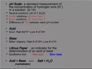
Slide31Buffers Keep the balance Buffers Keep the balance Acidosis- When blood pH drops below 7.35. Acidosis- When blood pH drops below 7.35. Alkalosis – When blood pH rises above 7.45. Alkalosis – When blood pH rises above 7.45. Sodium Bicarbonate ions NaHCO 3- Sodium Bicarbonate ions NaHCO 3- Bicarbonate ion HCO 3- Bicarbonate ion HCO 3- Ammonia NH 3 Ammonia NH 3 HCl HCl
Slide32Renal threshold Renal threshold The limit for certain substances in the blood. The limit for certain substances in the blood. Substances over the limit are forced out into the filtrate. Substances over the limit are forced out into the filtrate. Glucose and not reabsorbed over the threshold limit. Glucose and not reabsorbed over the threshold limit.
Slide33Renal Calculi AKA Kidney stones Renal Calculi AKA Kidney stones
Slide34Renal and Urinary disorders Renal and Urinary disorders Hydronephrosis – Urine backs up into the kidney, causing swelling. Hydronephrosis – Urine backs up into the kidney, causing swelling. Renal calculi – crystallized mineral chunks that develop in the calyces or renal pelvis(staghorn calculi) = renal colic(PAIN) Renal calculi – crystallized mineral chunks that develop in the calyces or renal pelvis(staghorn calculi) = renal colic(PAIN) Renal ptosis – Kidneys may drop, Ureters may kink and obstruct urine flow. Renal ptosis – Kidneys may drop, Ureters may kink and obstruct urine flow.
Slide35Urinary tract infections UTIs Urinary tract infections UTIs Urethritis -Bacterial infections – inflammation of the urethra- STDs often cause urethritis – more common in males Urethritis -Bacterial infections – inflammation of the urethra- STDs often cause urethritis – more common in males Cystitis – inflammation of the bladder- can also accompany kidney stones and tumors Cystitis – inflammation of the bladder- can also accompany kidney stones and tumors Nephritis – Kidney disease – bacterial or viral infections Nephritis – Kidney disease – bacterial or viral infections
Slide36Kidney Failure AKA Renal Failure Kidney Failure AKA Renal Failure Acute Renal Failure – Sudden Acute Renal Failure – Sudden BUN Blood Urea Nitrogen High BUN Blood Urea Nitrogen High 1 st stage Loss of nephrons 1 st stage Loss of nephrons 2 nd stage Kidney can no longer adapt to the loss of nephrons. Remaining nephrons cannot handle the load 2 nd stage Kidney can no longer adapt to the loss of nephrons. Remaining nephrons cannot handle the load 3 rd stage Complete shutdown- Edema and hypertension occurs 3 rd stage Complete shutdown- Edema and hypertension occurs