Understanding Cell Life: Structure, Transport, and Homeostasis

In Chapter 7 of this textbook, the focus is on the fundamental unit of life: the cell. The chapter covers the concept of life being cellular, and dives
- Uploaded on | 0 Views
-
 solan
solan
About Understanding Cell Life: Structure, Transport, and Homeostasis
PowerPoint presentation about 'Understanding Cell Life: Structure, Transport, and Homeostasis'. This presentation describes the topic on In Chapter 7 of this textbook, the focus is on the fundamental unit of life: the cell. The chapter covers the concept of life being cellular, and dives. The key topics included in this slideshow are . Download this presentation absolutely free.
Presentation Transcript
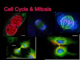
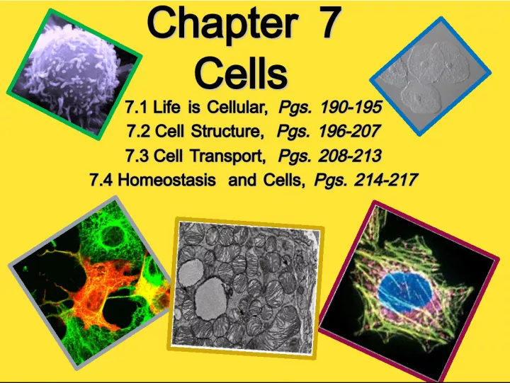
Slide1Chapter 7 Cells Chapter 7 Cells 7.1 Life is Cellular, Pgs. 190-195 7.1 Life is Cellular, Pgs. 190-195 7.2 Cell Structure, Pgs. 196-207 7.2 Cell Structure, Pgs. 196-207 7.3 Cell Transport, Pgs. 208-213 7.3 Cell Transport, Pgs. 208-213 7.4 Homeostasis and Cells, Pgs. 214-217 7.4 Homeostasis and Cells, Pgs. 214-217
Slide2Chapter Vocabulary7.1 Cell Cell Theory Cell Membrane Nucleus Eukaryote Prokaryote Cytoplasm Organelle Vacuole 7.2 Lysosome Cytoskeleton Centriole Ribosome Endoplasmic reticulum Golgi apparatus Chloroplast Mitochondrion Cell Wall Lipid Bilayer Selectively permeable 7.3 Diffusion Facilitated diffusion Aquaporin Osmosis Isotonic Hypotonic Hypertonic Osmotic pressure 7.4 Homeostasis Tissue Organ Organ system receptor
Slide37.1 Life is Cellular, Pgs. 190-195 7.1 Life is Cellular, Pgs. 190-195 • light microscopes can produce clear images of objects only to a magnification of about 1000 times. • Most living cells are nearly transparent, making it difficult to see the structures within them. • Using chemical stains or dyes can usually solve this problem. Some of these stains are so specific that they reveal only compounds or structures within the cell. f microscope Compound Light Microscopes and Cell Stains Onion skin cells
Slide4Light Microscopes and Cell Stains Human Cheek Cells **Can you see the nucleus inside? http://www.youtube.com/watch?v=7pR7TNzJ_pA&feature=related You can also hook the microscope up to a video camera and either a computer or tv. See the link below for a video taken of a light microscope slide containing an amoeba (a single-celled organism).
Slide5Electron Microscopes (SEM)• In scanning electron microscopes, a pencil-like beam of electrons is scanned over the surface of a specimen. • Because the image is of the surface, specimens viewed under a scanning electron microscope do not have to be cut into thin slices to be seen. • Scanning electron microscopes produce three- dimensional images of the specimen’s surface. Microbe Cholera Bacteria
Slide6Electron Microscopes (TEM) • Transmission electron microscopes make it possible to explore cell structures and large protein molecules. Because beams of electrons can only pass through thin samples, cells and tissues must be cut first into ultra thin slices before they can be examined under a transmission electron microscope. Thus, electron microscopy can be used to examine only nonliving cells and tissues. • Transmission electron microscopes produce flat, two-dimensional images. Mitochondrion from human lu ng
Slide7Early Contributions• Robert Hooke - First person to see cells, he was looking at cork and noted that he saw "a great many boxes. (1665) • Anton van Leeuwenhoek - Observed living cells in pond water, which he called "animalcules" (1673) Cork cells under The microscope Robert Hooke Anton van Leeuwenhoek
Slide8•Theodore Schwann - zoologist who observed tissues of animals had cells (1839) • Mattias Schleiden - botanist, observed tissues of plants contained cells ( 1845) • Rudolf Virchow - also reported that every living thing is made of up vital units, known as cells. He predicted that cells come from other cells . (1850 )
Slide9The Cell Theory• 1. Every living organism is made of one or more cells. • 2. The cell is the basic unit of structure and function. It is the smallest unit that can perform life functions. • 3. All cells arise from pre-existing cells. *Why is the Cell Theory called a Theory and not a Fact?
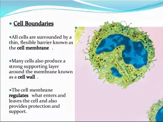
Slide10Cell FeaturesALL cell have these parts: • Ribosomes – make protein for use by the organism • Cytoplasm – fluid material within cell • DNA – genetic material • Cytoskeleton – internal framework of cell • Cell Membrane – outer boundary, some things can cross the cell membrane
Slide11Prokaryote Cells• The first cells to inhabit the earth • Simple cells • Bacteria • These cells do NOT have a nucleus, their DNA is circular and floats in the cytoplasm • Typical bacteria structure • Notice that there is nucleus inside.
Slide12Eukaryotic Cells• Cells found in plants, animals, protists, and fungi • 4 main parts: 1. Cell membrane 2. Cytoplasm 3. Nucleus 4. Organelles
Slide13•Usually found at center of cell • Has a nuclear membrane & nuclear pores • Contains cell’s DNA in one of 2 forms o chromatin - DNA bound to protein (non-dividing cell) o chromosomes - condensed structures seen in dividing cell • Also contains an organelle called nucleolus - which makes the cell’s ribosomes Section 7.2: Cell Structure, Pages 196-207 Nucleus
Slide14Mitochondria – this is the cell’s energy center. It turns food into a chemical energy called ATP The mitochondria is sometimes called the “powerhouse” of the cell
Slide15Golgi Apparatus – processes, packages and secretes proteins. It is comparable to a factory or a post office. *A vesicle forms with Golgi to transport substances outside cell.
Slide16Lysosome – Contains digestive enzymes, breaks things down, "suicide sac” Endoplasmic Reticulum – Transport, "intracellular highway". - Rough ER contains many ribosomes & is involves in protein synthesis - Smooth ER ribosomes not found on surface
Slide17Cytoskeleton – Helps cell maintain support & shape; movement a. microtubules -hollow structures; also help build cilia flagella b. microfilaments -threadlike c. centrioloes -only in animal cells; used during cell division (paired) Vacuole – storage area for water and other substaces, plant cells usually have a large central vacuole
Slide18THE ANIMAL CELL
Slide19THE PLANT CELL
Slide20Plant Cell PartsPlants have additional structures: • CELL WALL – surrounds membrane & provides additional support • CHLOROPLASTS – contain green pigment, function in photosynthesis • CENTRAL VACUOLE – large water container in center of cell
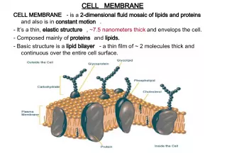
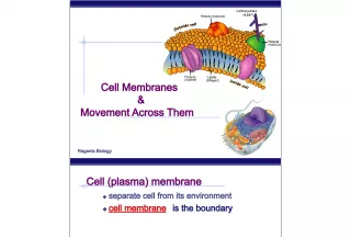
Slide21CELL MEMBRANE• It is composed of a double layer of phospholipids with proteins embedded throughout http://www.youtube.com/watch?v=moPJkCbKjBs&featur e=related
Slide22 A phospholipidAn animal cell Cell Membrane Composition 4 3 2 1
Slide23•The cell membrane is selectively permeable. In fact, this is one of the most important properties of the membrane. • What do you think selective permeability means? Maybe this picture will help. Selective Permeability Plasma membrane of budding yeast stained with green fluorescent dye
Slide24If you are still stuck, maybe this will helpThese objects are selectively permeable.
Slide257.3 Cell Transport, Pgs. 208-213Diffussion This is a type of passive transport because it does not use the cell’s energy (ATP). • The process by which particles move from an area of high concentration to an area of lower concentration is known as diffusion . http://www.indiana.edu/~phys215/lecture/lecnotes/lecgraphics/diffusion.gif • Diffusion is the driving force behind the movement of many substances across the cell membrane. http://www.youtube.com/watch?v=H7QsDs8ZRMI
Slide26Diffusion Diffusion depends upon random particle movements. http://www.youtube.com/watch?v=AYNwynwaALo&feature=related http://www.youtube.com/watch?v=s0p 1ztrbXPY&feature=related
Slide27Facilitated Diffusion- An example is OsmosisNo Energy Required!!!! • Molecules that cannot directly diffuse across the membrane pass through special protein channels in a process known as facilitated diffusion. • The movement of molecules by facilitated diffusion does not require any additional use of the cell’s energy. Can you see the channels in the membrane?
Slide28Osmosis: An Example of FacilitatedDiffusion • Many cells contain water channel proteins, known as aquaporins, that allow water to pass right through them. Without aquaporins, water would diffuse in and out of cells very slowly. *** Osmosis is the diffusion of water through a selectively permeable membrane.
Slide29Osmotic Pressure• For organisms to survive, they must have a way to balance the intake and loss of water. • The net movement of water out of or into a cell exerts a force known as osmotic pressure. http://www.youtube.com/watch?v=0c8acU E9Itw&feature=related http://www.youtube.com/watch?v=aeL6VL4 cAmE&feature=related Osmositic pressure causes water to move into or out of an egg
Slide30Osmotic Pressure• Cells placed in an isotonic solution have the same concentration of solution inside and outside of the cell. The shape of the cell does not change when placed in isotonic solution. • In a hypertonic solution, water rushes out of the cell, causing animal cells to shrink and plant cell vacuoles to collapse. • In a hypotonic solution, water rushes into the cell, causing cells to swell. Red Bood Cells in 3 Types of Media/Solutions Can you tell which solution is which?
Slide31Active Transport- Requires Energy• Cells sometimes must move materials against a concentration difference. • The movement of material against a concentration difference is known as active transport. Active transport requires energy. http://www.youtube.com/watch?v=STzOiR qzzL4&NR=1
Slide32Active Transport of Large Molecules• Larger molecules and clumps of material can also be actively transported across the cell membrane by processes known as endocytosis and exocytosis. http://www.youtube.com/watch?v=kfy 92hdaAH0&feature=channel http://www.youtube.com/watch?v=4gLt k8Yc1Zc&feature=related
Slide337.4 Homeostasis and Cells Pages 214-217• A single-celled, or unicellular, organism does everything you would expect a living thing to do. http://101science.com/paramecium.htm • Just like other living things, unicellular organisms must achieve homeostasis , relatively constant internal physical and chemical conditions. • To maintain homeostasis, unicellular organisms grow, respond to the environment, transform energy, and reproduce. The Cell as an Organism
Slide34Cell Specialization• The cells of multicellular organisms are specialized, with different cell types playing different roles. • Some cells are specialized to move, others to react to the environment, and still others to produce substances that the organism needs. • No matter what the role, each specialized cell contributes to the overall homeostasis of the organism. http://www.youtube.com/watch?v=9 duvzqvVflw • This bacteria is has specialized cilia to help it move. • What type of microscope was used here? How do you know?
Slide35Cellular Communication• Cells in a large organism communicate by means of chemical signals that are passed from one cell to another. • These cellular signals can speed up or slow down the activities of the cells that receive them, http://www.youtube.com/watch?v=ONljvPh 1ykY&feature=related • These cells are called neurons. They reach out and connect to other neurons so they can communicate with each other.