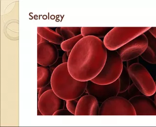
Characteristics of Blood

Blood is the only fluid connective tissue in the human body. It has an opaque, metallic taste and its color varies from scarlet to dull red depending on the level of oxygen it contains. Its pH ranges
- Uploaded on | 24 Views
-
 tristanhansen
About Characteristics of Blood
PowerPoint presentation about 'Characteristics of Blood'. This presentation describes the topic on Blood is the only fluid connective tissue in the human body. It has an opaque, metallic taste and its color varies from scarlet to dull red depending on the level of oxygen it contains. Its pH ranges. The key topics included in this slideshow are . Download this presentation absolutely free.
Presentation Transcript
Slide1Chapter 10BLOOD
Slide2Characteristics Only fluid (connective) tissue. Opaque Metallic taste Color – scarlet to dull red Depends on oxygen pH btw 7.35 to 7.45 temp 100.4 F (38C) higher than normal body temp 8% of body weight About 5-6 liters or 6 quarts
Slide3Formed Elements Erythrocytes – red blood cells (RBCs) 45% of blood Anucleate – no nucleus Contain hemoglobin molecules Iron bearing protein Transports most of the oxygen 1RBC = 250 million hmg molecules 1hmg can hold 4 oxygen molecules Biconcave discs About 5 million cells/mm3 More/less RBCs causes blood to thicken/thin Form hematocrit of spun blood
Slide4Formed elements cont. Anemia – decrease in oxygen carrying ability Sickle cell anemia – abnormal hmg causes RBC to become spiky and rupture easily Aplastic anemia – destruction of bone marrow by cancer Iron-deficiency anemia – lack of iron in diet or slow/prolonged bleeding Decrease in RBC # Pernicious anemia – lack of vitamin B12 Hemorrhagic anemia – hemorrhage Hemolytic anemia – bacterial infection causes lysis of RBCs
Slide5Polycythemia – increase in # of RBCs Causes Bone marrow cancer Living at high altitudes Thicker blood may move sluggishly and impair circulation
Slide6Formed elements cont. Leukocytes – white blood cells (WBCs) 4000 to 11000/mm3 < 1% of total blood volume Are complete cells Diapedesis – WBCs can move in/out of vessels to reach infected/inflamed areas Positive chemotaxis – WBCs respond to chemicals released by damaged cells
Slide7Formed elements cont. When needed their # doubles >11000 = leukocytosis Indicates an infection <4000 = leukopenia Caused by some drugs – corticosteroids and anticancer agents Leukemia – bone marrow produces large #s of WBCs that are unable to function
Slide8Formed elements cont. 2 groups – based on visible granules Granulocytes – lobed nuclei Neutrophils – multilobed nuclei o Most numerous/phagocytic o Fine granules o @ sites of acute infections Eosinophils – blue-red nucleus o Large granules o Respond to allergies and infections by worms Basophils – rarest o U or S shaped nuclei o Granules w/ histamine – attracts other WBCs to inflamed site
Slide9Formed elements cont. Agranulocytes – normal nuclei Lymphocytes o Large purple nucleus o In lymphatic tissue o Immune system Monocytes – largest o Change into macrophages o Fight chronic infections
Slide10Formed elements cont. Platelets – fragments of multinucleate cells called megakaryocytes 300,000/mm3 Blood clotting Cling to broken area
Slide11Plasma Nonliving fluid matrix 55% of blood 90% water Straw color Includes nutrients, salts, gases, hormones, wastes, and plasma proteins Plasma proteins Produced by liver Albumin – contributes to osmotic pressure Keeps water in bloodstream Clotting proteins Antibodies – protect from pathogens
Slide12Hematopoiesis – blood cell formation Occurs in myeloid tissue (red bone marrow) Hemocytoblast – stem cell in marrow that produces formed elements Forms 2 types of secondary stem cells Lymphoid stem cells – produce lymphocytes Myeloid stem cells – produce erythrocytes and platelets Fully formed RBCs do not grow or divide Life span btw 100-120 days Become rigid and break apart Spleen and liver get rid of pieces
Slide13Hematopoiesis cont. RBC Development – 3-5 days Young RBCs divide until enough hmg is produced Then eject nucleus and all organelles except ER Called a reticulocyte Enter bloodstream w/in 2 days of release eject ER Become erythrocytes Erythropoietin – hormone – controls production rate Produced at a constant rate by kidneys Decrease in oxygen = increase in production
Slide14Hematopoiesis cont. WBCs – production stimulated by colony stimulating factors (CSFs) and interleukins Platelets – production stimulated by thrombopoietin Bone marrow biopsy – taking a sample for examination
Slide15Maintaining hemostasis (stoppage ofblood flow) Occurs when vessel walls break 3 phases Platelet plug forms – aka white thrombus Platelets stick to damaged site Vascular spasms occur Platelets release serotonin – causes spasms Slow blood loss until clot forms Coagulation events occur Tissues release tissue factor (TF) PF3 (on platelets) reacts w/ TF, vitamin K, and Ca+2 to trigger clotting cascade Prothrombin activator converts prothrombin in plasma to thrombin Thrombin joins fibrinogen to form fibrin (mesh) to capture RBCs
Slide16Hemostasis cont. Usually takes w/in 3-6 minutes Disorders Undesirable clotting Thrombus – clot in an unbroken vessel May cause blockage Embolus – thrombus that floats in bloodstream May move to brain – stroke Bleeding disorders Thrombocytopenia – insufficient # of platelets Hemophilia – genetic – lack any factors needed for clotting
Slide17Human Blood Groups Plasma membranes of RBCs have proteins (cellular or self antigens) Antigens – substances the body recognizes as foreign Stimulate the immune system to release antibodies Antibodies in plasma attach to surface antigens different from those of the individual Agglutination – clumping of cells due to antibodies Leads to clogging of vessels and lysis of foreign cells Lysis releases hmg which may clog kidney tubules = kidney failure
Slide18Blood groups cont. ABO blood group Based on 2 antigens – type A or type B Absence of both = type O – universal donor Presence of both = type AB – universal recipient From birth your body contains antibodies for the ABO antigens not present – anti-A or anti-B antibodies
Slide19Blood groups cont. Rh blood group Rh+ = Rh antigen is present Rh- = Rh antigen is absent anti-Rh antibodies do not form until exposed to Rh+ blood Rh- women have to be careful when pregnant 1 st Rh+ baby – health 2 nd Rh+ baby – mother’s body now has antibodies and will attempt to destroy RBCs of the baby = hemolytic disease of the newborn May prevent by taking RhoGAM shortly after the birth of her 1 st Rh+ baby – prevents production of antibodies
Slide20Blood Typing Test blood by mixing with anti-A and anti-B immune serum. Agglutination will occur btw the antibody and the corresponding antigen (if present). Type AB – contains antigens A and B – will agglutinate with both antibodies Type B – contains antigen B – agglutinates with anti-B Type A – contains antigen A – agglutinates with anti-A Type O – no antigens – no agglutination occurs
Slide21Development Embryonic blood cells are circulating by day 28 Fetal hemoglobin (HbF) is present – can carry more oxygen At birth fetal cells are broken down If broken down too fast for liver – physiologic jaundice may occur