Consensus on diagnosis and treatment of superficial thrombophlebitis

Establishing a uniform approach to diagnosing and treating superficial thrombophlebitis will ensure consistent and effective care across medical specialties.
- Uploaded on | 1 Views
-
 carolyn
About Consensus on diagnosis and treatment of superficial thrombophlebitis
PowerPoint presentation about 'Consensus on diagnosis and treatment of superficial thrombophlebitis'. This presentation describes the topic on Establishing a uniform approach to diagnosing and treating superficial thrombophlebitis will ensure consistent and effective care across medical specialties.. The key topics included in this slideshow are . Download this presentation absolutely free.
Presentation Transcript
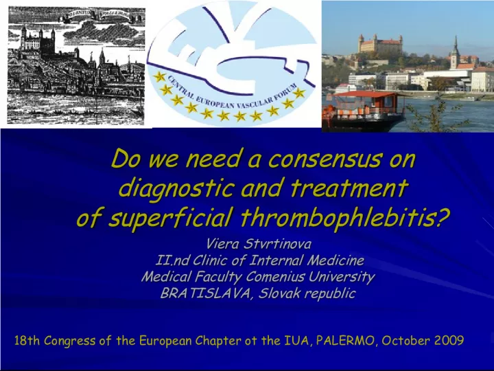
1. Do we need a consensus on diagnostic and treatment of superficial thrombophlebitis? Do we need a consensus on diagnostic and treatment of superficial thrombophlebitis? Viera Stvrtinova Viera Stvrtinova II.nd Clinic of Internal Medicine II.nd Clinic of Internal Medicine Medical Faculty Comenius University Medical Faculty Comenius University BRATISLAVA, Slovak republic BRATISLAVA, Slovak republic 18th Congress of the European Chapter ot the IUA, PALERMO, October 2009
2. INTRODUCTION INTRODUCTION • Traditionally ST has been considered a relatively benign and limited disease or sign of chronic venous insufficiency • In recent years it seems that ST is not such a banal condition • S ome physicians consider ST an integral part of venous thromboembolism, together with DVT an d PE
3. ST and VTE ST and VTE • Association between ST and DVT vary in the literature from 6 to 53% and pulmonary embolism up to 33% • The relationship between ST and DVT is supported also by the same risk factors that triggers ST as well as DVT.
4. in ST a DVT – always other part of Virchow´s triad is more important in ST a DVT – always other part of Virchow´s triad is more important Rudolf VIRCHOW (1821-1902)
5. RISK FACTORS for ST RISK FACTORS for ST • Slowing of the blood flow • Slowing of the blood flow • Varicose veins • Varicose veins • Prolonged bed rest (immobilization) for several reasons (operation, trauma, serious infection, heart failure, pulmonary emphysema, etc.) • Prolonged bed rest (immobilization) for several reasons (operation, trauma, serious infection, heart failure, pulmonary emphysema, etc.)
6. RISK FACTORS for ST RISK FACTORS for ST • Coagulation disorders • Coagulation disorders • Thrombophilia – Factor V Leiden mutation, protein C deficiency, protein S deficiency, AT III deficiency, prothrombin G20210A mutation, etc • Thrombophilia – Factor V Leiden mutation, protein C deficiency, protein S deficiency, AT III deficiency, prothrombin G20210A mutation, etc • Cancer • Cancer • Pregnancy • Pregnancy • Oral contraceptives • Oral contraceptives • Anti-phospholipid sy • Anti-phospholipid sy
7. RISK FACTORS for ST RISK FACTORS for ST • Damage of the venous wall • Damage of the venous wall • Intravenous injections • Intravenous injections • Intravenous catheters • Intravenous catheters • Injury, trauma • Injury, trauma • Varicose veins and chronic venous insufficiency • Varicose veins and chronic venous insufficiency
8. RISK FACTORS for ST RISK FACTORS for ST • Other risk factors and diseases • Other risk factors and diseases • Winiwarter-Buerger´s disease • Winiwarter-Buerger´s disease • Behcet´s disease • Behcet´s disease • Other chronic inflammatory autoimmune diseases • Other chronic inflammatory autoimmune diseases • Obesity • Obesity • Age (over 60 yrs) • Age (over 60 yrs)
9. ST - incidence ST - incidence • S T is a frequent disease, but its exact incidence is not known. • The incidence of ST could be around 400 cases in 100.000 person – years according to UK estimates • It is depending on the age of the population, on the used investigation method (clinical dg. or DUS), and the fact, that ST is mostly managed on GPs level
10. ST - Classification ST - Classification According the etiology: 1.PRIMARY ST (inflammation affects only the venous wall and surrounding peri-venous tissue) 2.SECONDARY ST (inflammation of the venous wall is associated with other inflammatory process or systemic disease in the body According the etiology: 1.PRIMARY ST (inflammation affects only the venous wall and surrounding peri-venous tissue) 2.SECONDARY ST (inflammation of the venous wall is associated with other inflammatory process or systemic disease in the body
11. Primary ST Primary ST 1. varicophlebitis 1. varicophlebitis 2. septic catheter´s ST (e.g. non sterile i.v. injections in drug abusers, i.v.catheters in patients with immunodeficiency ) 2. septic catheter´s ST (e.g. non sterile i.v. injections in drug abusers, i.v.catheters in patients with immunodeficiency ) 3. “sterile” ST due to intravenous drug administration (scleroterapy) 3. “sterile” ST due to intravenous drug administration (scleroterapy) 4. Mondor thrombophlebitis 4. Mondor thrombophlebitis
12. Secondary ST Secondary ST 1. Winiwarter – Buerger´s disease 1. Winiwarter – Buerger´s disease 2. Behcet´s disease 2. Behcet´s disease 3. other vasculitic or chronic rheumatic syndromes with autoimmune etiology (e.g.anti-phospolipid syndrome) 3. other vasculitic or chronic rheumatic syndromes with autoimmune etiology (e.g.anti-phospolipid syndrome) 4. malignant tumors 4. malignant tumors
13. FORMS of ST FORMS of ST 1.varicophlebitis – ST of a varicose vein (VST) 1.varicophlebitis – ST of a varicose vein (VST) 2 . ST on a healthy, non-varicose superficial vein – non-varicose ST (NVST) 2 . ST on a healthy, non-varicose superficial vein – non-varicose ST (NVST) 88% of ST 88% of ST are VST are VST
14. Endothelial injury in NVST Circulatory stasis in Circulatory stasis in varicophlebitis always other part of Virchow´s triad is more important always other part of Virchow´s triad is more important
15. Varicophlebitis and DVT Varicophlebitis and DVT In a retrospective study of 114 patients with ST the incidence of a concomitant DVT was 15.6% when ST affected the vena saphena magna or vena saphena parva, but only 5.2% when side branches were involved. In a retrospective study of 114 patients with ST the incidence of a concomitant DVT was 15.6% when ST affected the vena saphena magna or vena saphena parva, but only 5.2% when side branches were involved. With varicose veins as a single risk factor, the frequency of a concomitant DVT was 6%, varicose veins combined with further risk factors showed a DVT frequency of 15.4% (Noppeney et al, 2006). With varicose veins as a single risk factor, the frequency of a concomitant DVT was 6%, varicose veins combined with further risk factors showed a DVT frequency of 15.4% (Noppeney et al, 2006).
16. NVST (“non-varicose” superficial thrombophlebitis) NVST (“non-varicose” superficial thrombophlebitis) • is a miscellaneous group of disorders , where inflammation is a dominating feature in some conditions, while thrombosis dominates in other cases. • Among 2319 patients diagnosed with Behcet´s disease ST was present in 53.3% and DVT in 29,8% of cases
17. ST in Winiwarter´s – Buerger´s dis. ST in Winiwarter´s – Buerger´s dis. • ST in Buerger´s disease belongs to the diagnostic criteria • Thrombophlebitis migrans (inflammation of the venous wall goes up or down – proximally or distally on a superficial vein) or thrombophlebitis saltans (inflammation “jumps” from one vein to another vein) are specific forms of ST often seen in patients with Winiwarter- Buerger´s disease.
18. NVST NVST • In a prospective analysis of 42 patients with non varicose ST investigation for risk factors revealed a neoplasm in 2 patients (4.8%), a non neoplasic systemic disease in 4 (9.5%) a thrombophilic condition in 20 patients (48%). • The most frequent thrombophilia was the heterozygous mutation of coagulation factor V Leiden – ( Gillet et al, 2004).
19. ST diagnosis ST diagnosis • In many cases ST is a banal condition, which resolves spontaneously, but in recent years due to systematic ultrasound investigation of the venous system a large number of deep venous thromboses concomitant with ST has been revealed.
20. Thrombophlebitis Thrombophlebitis DUS
21. ST treatment ST treatment On contrary to the treatment of deep venous thrombosis, only little is known about the most appropriate management of ST. On contrary to the treatment of deep venous thrombosis, only little is known about the most appropriate management of ST. ST is etiologically a heterogeneous group of disorders with a different degree of inflammation and thrombosis ST is etiologically a heterogeneous group of disorders with a different degree of inflammation and thrombosis the main etiological factor and contribution of different risk factors always should be considered before treatment decision. the main etiological factor and contribution of different risk factors always should be considered before treatment decision.
22. ST th. – compression , mobilization ST th. – compression , mobilization The main therapeutic procedure in all types of ST is compression and mobilization. The main therapeutic procedure in all types of ST is compression and mobilization. There have been no randomized studies demonstrating the effectiveness of compression, although this approach is considered by all experts to be essential. There have been no randomized studies demonstrating the effectiveness of compression, although this approach is considered by all experts to be essential. In all cases of ST immediate mobilization with elastic compression is necessary. In all cases of ST immediate mobilization with elastic compression is necessary.
23. All patients with ST s hould be treated with compression therapy. All patients with ST s hould be treated with compression therapy. Regul ar walking supports the effectiveness of the compression bandage on the LL . The patient must walk regularly throughout the day and avoid prolonged periods of being seated or standing. Confinement to bed would favor progression of the thrombus in both the superficial and the deep venous system Regul ar walking supports the effectiveness of the compression bandage on the LL . The patient must walk regularly throughout the day and avoid prolonged periods of being seated or standing. Confinement to bed would favor progression of the thrombus in both the superficial and the deep venous system
24. ST treatment - DRUGS ST treatment - DRUGS Anticoagulants Anticoagulants Non-steroidal anti-inflammatory drugs (NSAIDs) Non-steroidal anti-inflammatory drugs (NSAIDs) Topical local anti-inflammatory treatment (gel, cream, spray) Topical local anti-inflammatory treatment (gel, cream, spray) Venoactive drugs – in patients with varicose ST Venoactive drugs – in patients with varicose ST Antibiotics – in patients with septic ST Antibiotics – in patients with septic ST Corticosteroids – in patients with vasculitic and autoimmune syndromes Corticosteroids – in patients with vasculitic and autoimmune syndromes
25. ST treatment - anticoagulants ST treatment - anticoagulants Especially in cases of extensive ST anticoagulant therapy is a good choice. LWMH, UFH as well as oral anticoagulants are used in prophylactic as well as therapeutic doses. Especially in cases of extensive ST anticoagulant therapy is a good choice. LWMH, UFH as well as oral anticoagulants are used in prophylactic as well as therapeutic doses. Not only the doses, but the duration of the treatment is different in individual hospitals and medical care centers. Not only the doses, but the duration of the treatment is different in individual hospitals and medical care centers.
26. LMWH and ST LMWH and ST ? ? ? ? ? ? ? ? ? ? ? ? ? ? Dosage – prophylactic or therapeutic Dosage – prophylactic or therapeutic Duration – 10, 20, 30 days ???????? Duration – 10, 20, 30 days ????????
27. Proposal for ST management from CEVF Proposal for ST management from CEVF Recommendation n.1: Recommendation n.1: In every patient with NVST and in every patient with recurrent VST look carefully for risk factors for superficial thrombophlebitis, especially for thrombophilia and cancer In every patient with NVST and in every patient with recurrent VST look carefully for risk factors for superficial thrombophlebitis, especially for thrombophilia and cancer 18th Congress of the European Chapter ot the IUA, PALERMO, October 2009
28. Recommendation n.2: Recommendation n.2: Clinical investigation may the real extent of superficial thrombophlebitis underestimate, and does not give enough information on the status of deep venous system, therefore after clinical investigation it is important to perform duplex ultrasound investigation of the superficial and deep venous system, too. Clinical investigation may the real extent of superficial thrombophlebitis underestimate, and does not give enough information on the status of deep venous system, therefore after clinical investigation it is important to perform duplex ultrasound investigation of the superficial and deep venous system, too. 18th Congress of the European Chapter ot the IUA, PALERMO, October 2009
29. Recommendation n.3: Recommendation n.3: Duplex ultrasound investigation should be done bilaterally - on both lower limbs, not only on the limb affected with ST. Duplex ultrasound investigation should be done bilaterally - on both lower limbs, not only on the limb affected with ST. 18th Congress of the European Chapter ot the IUA, PALERMO, October 2009
30. It is necessary (mandatory) to perform duplex ultrasound investigation immediately after clinical diagnosis of ST in the case of ST localized on the trunk of the great saphenous vein 10 cm and less from the sapheno-femoral junction or on the trunk of small saphenous vein 10 cm or less from the sapheno-popliteal junction. It is necessary (mandatory) to perform duplex ultrasound investigation immediately after clinical diagnosis of ST in the case of ST localized on the trunk of the great saphenous vein 10 cm and less from the sapheno-femoral junction or on the trunk of small saphenous vein 10 cm or less from the sapheno-popliteal junction. Recommendation n.4: Recommendation n.4: 18th Congress of the European Chapter ot the IUA, PALERMO, October 2009
31. Recommendation n.5: Recommendation n.5: All patients with superficial thrombophlebitis should be treated with compression therapy. All patients with superficial thrombophlebitis should be treated with compression therapy. Recommendation n.6: Recommendation n.6: In all cases of ST immediate mobilization with elastic compression is necessary (mandatory). Patients should not be confined to bed. In all cases of ST immediate mobilization with elastic compression is necessary (mandatory). Patients should not be confined to bed. Proposal for Proposal for ST management from CEVF ST management from CEVF 18th Congress of the European Chapter ot the IUA, PALERMO, October 2009
32. Patients with ST, with an inflamed and thrombosed superficial vein longer than 5 cm in duplex ultrasound investigation should have anticoagulant treatment with LMWH for 4 weeks. The dosage and duration of anticoagulation depends on the concomitant diseases and other risk factors for VTE. Patients with ST, with an inflamed and thrombosed superficial vein longer than 5 cm in duplex ultrasound investigation should have anticoagulant treatment with LMWH for 4 weeks. The dosage and duration of anticoagulation depends on the concomitant diseases and other risk factors for VTE. Recommendation n.7: Recommendation n.7: 18th Congress of the European Chapter ot the IUA, PALERMO, October 2009
33. In conclusion In conclusion Do we need a consensus on diagnostic and treatment of superficial thrombophlebitis? Do we need a consensus on diagnostic and treatment of superficial thrombophlebitis?