Tetralogy of Fallot: Anatomy, Pathophysiology and History

This article details the anatomy, pathophysiology and history of Tetralogy of Fallot, a congenital heart defect. It covers important milestones in its discovery, and the underlying anatomic features that cause the disease.
- Uploaded on | 4 Views
-
 masamismith
About Tetralogy of Fallot: Anatomy, Pathophysiology and History
PowerPoint presentation about 'Tetralogy of Fallot: Anatomy, Pathophysiology and History'. This presentation describes the topic on This article details the anatomy, pathophysiology and history of Tetralogy of Fallot, a congenital heart defect. It covers important milestones in its discovery, and the underlying anatomic features that cause the disease.. The key topics included in this slideshow are Tetralogy of Fallot, congenital heart defect, anatomy, pathophysiology, history,. Download this presentation absolutely free.
Presentation Transcript
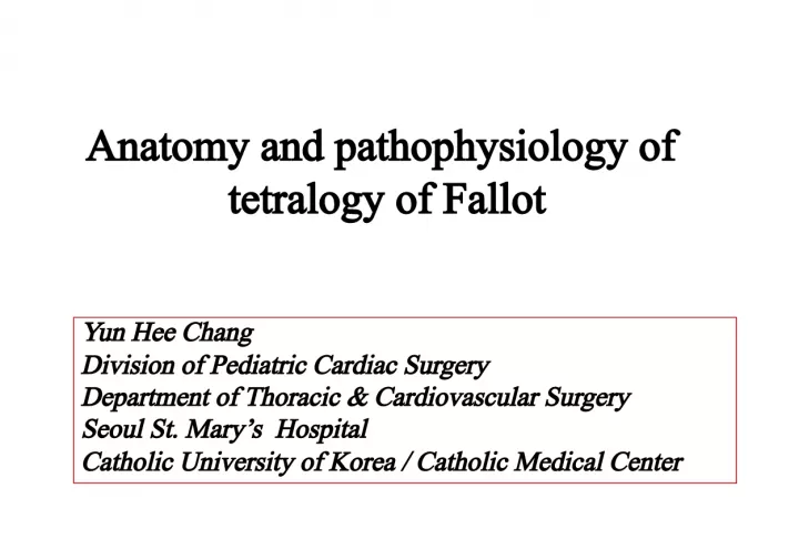
1. Anatomy and pathophysiology of tetralogy of Fallot Yun Hee Chang Division of Pediatric Cardiac Surgery Department of Thoracic & Cardiovascular Surgery Seoul St. Marys Hospital Catholic University of Korea / Catholic Medical Center
2. History
3. 1671 1777 1785 1793 1797 1812 1814 1816 1846 1881 1888 Niels Stensen Eduard Sandifort Willam Hunter Pulteney 1784 Abernethy Bell Dorsey J.P.Farre Thaxter Thomas Bevil Peacock Widman Fallot: La maladie bleue 1924 Maude Abbott : tetralogy of Fallot 4 Anatomic features (1) Pulmonary (or RV) outflow stenosis (2) Ventricular septal defect (3) Aortic overriding (4) Right ventricular hypertrophy
4. Subtypes of TOF Subtypes of TOF Tetralogy of Fallot, Pulmonary stenosis Tetralogy of Fallot, Absent pulmonary valve (3-6%) Tetralogy of Fallot, Common atrioventricular canal (2%) Tetralogy of Fallot, Pulmonary atresia (20%)
5. TOF with Pulmonary stenosis
6. Anatomic features Anatomic features Overriding of the aorta Subpulmonary stenosis Ventricular septal defect Right ventricular hypertrophy
7. Van Praagh R et al. - Underdevelopment of the subpulmonary infundibulum Normal TOF Pathognomonic lesions Pathognomonic lesions
8. Anderson RH et al. - Anterocephalad deviation of the outlet septum (relative to the limb of the septomarginal trabeculation) - Malformation of the septoparietal trabeculation Normal TOF A P
9. Deviated outlet septum Septal attachment of the muscular outlet septum Antero-cranial limb of TSM Hypertrophied Septoparietal trabeculation
10. Subvalvar stenosis Infundibular stenosis - Essential part of tetralogy - Produced by the Squeeze between the anterocephalad malalignment of the outlet septum and the abnormal situated septoparietal trabeculations Pulmonary outflow stenosis Pulmonary outflow stenosis
11. Anterocephalad malalignment of the outlet septum without abnormal situated septoparietal trabeculations * vs . Eisenmenger type ventricular septal defect
12. Additional muscular stenosis - By hypertrophy of the moderator band or by prominent apical trabeculations. - Often described as two-chambered right ventricle. Moderator band Apical trabeculations
13. - The pulmonary valve is stenotic in 75% cases : usually caused by hypoplasia and fusion of bicuspid leaflets, supravalvar tethering . - The valve is bicuspid in to 2/3 of cases. - The pulmonary valve annulus is invariably smaller than the aorta; however, it is not necessarily significant obstructive. V alvar stenosis
14. - The main PA is usually somewhat diffusely small and is often short. - The narrowed portion of the main pulmonary artery is often at the sinotubular junction . - Branch PA abnormalities occurred in only 10 % of cases. Sinotubular junction Suprav alvar stenosis
15. Outlet from left ventricle Interventricular plane Ventricular septal defect Ventricular septal defect Ventricular septal defect 3 important planes
16. Perimembranous defect - In about 4/5 of Caucasian patients - VIF stops short of the postero-caudal limb of TSM, permitting fibrous continuity to exist between the leaflets of the aortic & tricuspid valves Muscular outlet septum Ventricular infundibular fold Remnant of the interventricular membranous septum AV node Right bundle branch Left bundle branch Postero-caudal limb of TSM Types of ventricular septal defect
17. Ventricular infundibular fold Septomarginal trabeculation Septoparietal trabeculation Aortic-tricuspid continuity
18. Muscular defect - In about 1/5 of Caucasian patients - Postero-caudal limb of the TSM fuses with VIF, permitting muscular continuity throughout the right ventricular margin of the defect. Postero-caudal limb of TSM Ventricular infundibular fold Muscular outlet septum Hypertrophied septoparietal trabeculation Right bundle branch Left bundle branch AV node
19. Ventricular infundibular fold Septomarginal trabeculation Septoparietal trabeculation
20. Doubly committed & juxta-arterial defect - Commoner in the Far East and South America - Consequence of failure of formation of a complete muscular subpulmonary infundibulum Ventricular infundibular fold Fibrous continuity between the leaflets of the arterial valves Postero-caudal limb of TSM
21. Unobstructed but relatively high- resistance systemic vascular bed Obstructed pulmonary outflow tract Anatomic route of balance between the two circulatory beds RV hypertension Normal or low PA pressure Pathophysiology Pathophysiology
22. Hypercyanotic spell Catecholamine State of low intravascular volume state Crying /feeding, etc
23. TOF with Pulmonary atresia
24. Anatomic features Anatomic features
26. - Confluent or non-confluent . - Confluent in about 2/3 of the cases - The caliber of the central PAs varies : When the ductus or collateral arteries connect proximally to the central PAs or their lobar branches, the central vessels may be only mildly hypoplastic or even normal in size. Atretic arterial segment - Can be recognized as a solid elastic cord in about - Rarely only the PV is imperforate. The central right & left PAs
27. Entirely from the systemic circulation. - Ductus arteriosus - Systemic-to - pulmonary collateral arteries - Coronary artery - Plexus of bronchial or pleural arteries The blood supply to the lungs
28. Systemic-to-pulmonary collateral arteries Pulmonary arteries Pulmonary atreisia Patent ductus arteriosus * Ductal & collateral sources may coexist in the same patients but only rarely coexist in the same lung segment.
29. Ductus arteriosus - Usually is a unilateral structure - Associated with confluent PAs in > 80% of cases - Rarely, bilateral ductus may occur with non-confluent arteries - Because the ductus is widely patent during fetal life, the PAs may be a normal size at birth. - Normal postnatal ductal narrowing usually occurs and produce distal stenosis in 35-50% of cases. Patent ductus arteriosus Pulmonary coarctation
30. Collateral arteries - Most commonly from the descending thoracic aorta - Less commonly the subclavian arteries - Rarely from the abdominal aorta - Their number varies from 1 to 6 - Their diameter ranges from 1 to 20mm - More stable source of pulmonary blood flow
31. - Anastomoses between the central PAs (or their branches) and the collateral arteries : About 40% of subjects : May occur at the hilum or within the lung : In the remaining 60%, the collateral arteries enter the pulmonary hilum, travel with the bronchi as PAs
32. - Stenosis : nearly 60% of collateral arteries : tend to occur near the aortic or intrapulmonary anastomosis : may be discrete or segmental : may be congenital or acquired
33. - Ductus supplies confluent central PAs : Intra-PAs of both lungs are normal - Ductus supplies one of the non-confluent central PAs : Contra-lateral lung usually has arborization abnormalities - Ductus is absent : Both lungs have arborization abnormalities Intrapulmonary artery distribution
34. Classification Classification - There is no standard classification system for PAVSD, but several have been proposed. - Most classification schemes focus on the patterns of pulmonary blood flow. Congenital Heart Surgery Nomenclature and Database Project
35. Boston group IIIa. Central pulmonary artery Z score > -2.5 IIIb. Central pulmonary artery Z score < -2.5
36. Pathophysiology Pathophysiology One of Three Marked heart failure because of lung overflow Cyanotic because of reduced lung flow Fairly well balanced with systemic oxygen saturation in the high 70s to low 80s Extrapulmonary collateral Obstruction Collateral stenosis Intrapulmonary collateral has thin- walled elastic media
37. TOF with Absent Pulmonary Valve
38. Absence of ductus arteriosus Pulmonary annular hypoplasia Anatomic features Anatomic features Dilated pulmonary arterial trees Deviated muscular outlet septum Rudimentary leaflets of PV
39. Pathophysiology Pathophysiology Free pulmonary regurgitation throughout fetal life - Transmission of chronic volume load of the RV to PAs Proximal PA : aneurismal dilatation PA Normal TOF with APV Airway compression
40. Common atrioventricular canal
41. Anatomic features Anatomic features Septoparietal trabeculations Outlet septum Pulmonary trunk Aorta Anterior papillary muscle Common atrioventricular valve