From DNA to Proteins: Exploring Metabolic Pathways and Byssus in Mussels

In this chapter of the Honors Biology program at Mountain Pointe High School, students will delve into the fascinating world of metabolic pathways
- Uploaded on | 1 Views
-
 rafaeldelgado
About From DNA to Proteins: Exploring Metabolic Pathways and Byssus in Mussels
PowerPoint presentation about 'From DNA to Proteins: Exploring Metabolic Pathways and Byssus in Mussels'. This presentation describes the topic on In this chapter of the Honors Biology program at Mountain Pointe High School, students will delve into the fascinating world of metabolic pathways. The key topics included in this slideshow are . Download this presentation absolutely free.
Presentation Transcript
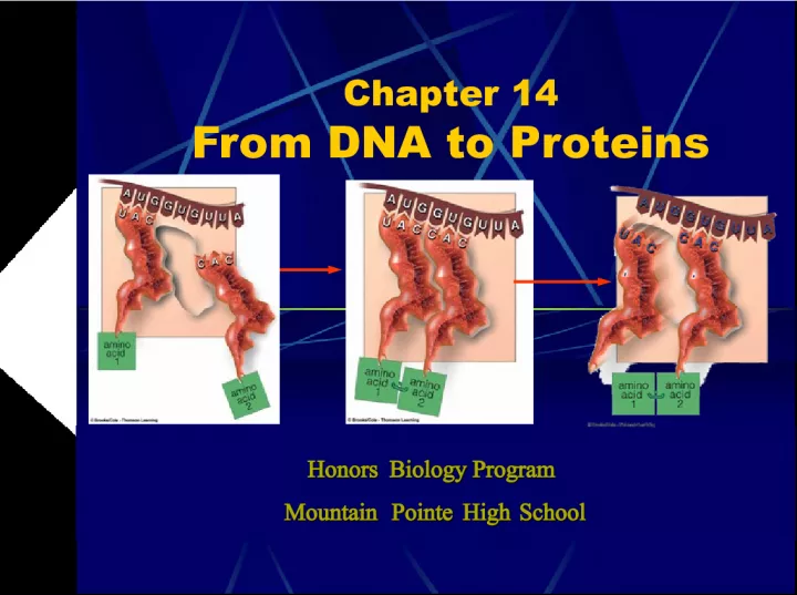
Slide1Chapter 14From DNA to Proteins Honors Biology Program Honors Biology Program Mountain Pointe High School Mountain Pointe High School
Slide2What are the byssus of a mussels?Why are these byssus so important?
Slide3Archibald GarrodSTEPS OF A METABOLIC PATHWAY: STEPS OF A METABOLIC PATHWAY: First to notice that many heritable diseases were related to metabolic pathway malfunctions. A B C X D Action of enzyme 1 Action of enzyme 2 Something has interfered with the action of enzyme 3. Completion of the pathway is blocked, and C accumulates. Garrod hypothesized that each of his affected patients had inherited a single metabolic defect that interfered with an enzyme in a particular metabolic pathway.
Slide4Beadle & Tatum33 years after Garrod’s hypothesis, these scientists were experimenting with a common bread mold that’s capable of synthesizing everything it needs to survive except for a few basic substances. Neurospora crassa and other fungal species They discovered that some of the fungal strains would only grow when supplied with vitamin B 6 , others would only grow in the presence of B 12 , etc.
Slide5Beadle & TatumAfter careful examination, they discovered that there was a different defective enzyme in each mutant strain of the fungus. In other words, each strain of fungus possessed an inherited mutation that corresponded to a defective enzyme. In other words, each strain of fungus possessed an inherited mutation that corresponded to a defective enzyme. This evidence supported Garrod’s “one gene, one enzyme” hypothesis! This evidence supported Garrod’s “one gene, one enzyme” hypothesis!
Slide6Sickle-cell AnemiaThe most common lethal genetic disease in African Americans, it causes normal red blood cells to become sickle shaped, which causes an incredible variety of health problems for its victims. Normal red blood cell Sickled red blood cell
Slide7Sickle-cell Anemiahemoglobin It was discovered that this disease was caused by a defect in a protein known as hemoglobin that is found in red blood cells. Normal hemoglobin is designated HbA . Normal hemoglobin is designated HbA . Abnormal hemoglobin is designated HbS . Abnormal hemoglobin is designated HbS .
Slide8Pauling &Itano gel electrophoresis In 1949, these scientists subjected molecules of HbA and HbS to gel electrophoresis . In this procedure, an electric field is used to move molecules through a gel. Molecules are separated by their size, shape & surface charge . upper buffer solution electrode glass tube or plates containing gel gel lower buffer solution power supply electrode movement of proteins
Slide9Pauling &Itano HbA HbA molecules carried the greatest surface charge and therefore moved through the gel the fastest. HbS HbS molecules moved much slower. As molecules move through the gel, they’re separated into distinct bands.
Slide10Vernon IngramHbA HbS Pinpointed the biochemical difference between HbA and HbS Hemoglobin (left) is a molecule made of 4 polypeptide chains , 2 alpha & 2 beta. Hemoglobin (left) is a molecule made of 4 polypeptide chains , 2 alpha & 2 beta. Ingram discovered that the defect was caused by an incorrect amino acid substitution in one of the beta chains! Ingram discovered that the defect was caused by an incorrect amino acid substitution in one of the beta chains!
Slide11```VALINE VALINE VALINE HISTIDINE HISTIDINE LEUCINE LEUCINE PROLINE PROLINE THREONINE THREONINE GLUTAMATE GLUTAMATE GLUTAMATE A beta chain Hemoglobin molecule Beta chain of an HbA molecule Beta chain of an HbS molecule
Slide12The discovery of the differencebetween the alpha & beta chains of hemoglobin meant that… Two genes must code for hemoglobin, one for each type of polypeptide chain. Genes code for all proteins , not just enzymes. The amino acid sequences of polypeptide chains are encoded in genes.
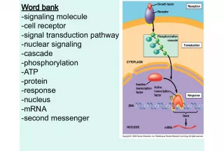
Slide133 Different Types of RNA
Slide14The Three Types of RNAmRNA protein-building instructions mRNA is a single-stranded molecule that takes DNA’s protein-building instructions out of the nucleus. rRNA ribosomes rRNA is the primary component of ribosomes , the organelles that actually make proteins. tRNA delivering amino acids tRNA is the molecule responsible for delivering amino acids one by one to a ribosome in the correct order specified by the mRNA molecule. mRNA A messenger RNA molecule ( mRNA ) rRNA A ribosomal RNA molecule ( rRNA ) tRNA A transfer RNA molecule ( tRNA )
Slide15Comparing DNA and RNADouble-stranded Deoxyribose sugars 4 nitrogenous bases Adenine Cytosine Guanine Thymine Single-stranded Ribose sugars 4 nitrogenous bases Adenine Cytosine Guanine Uracil Uracil Like thymine, uracil (at right, in blue) is a pyrimidine and is capable of pairing with adenine.
Slide16TranscriptionTranscription template mRNA Transcription is the process of using a portion of the DNA molecule as a template to assemble a molecule of mRNA . selected stretch of one DNA strand Only a selected stretch of one DNA strand is used as a template. promoter gene Transcription is initiated at a promoter , a DNA base sequence that signals the start of a gene . sugar-phosphate backbone of one strand of nucleotides in a DNA double helix sugar-phosphate backbone of the other strand of nucleotides part of the sequence of base pairs in DNA transcribed DNA winds up again DNA to be transcribed unwinds Newly forming RNA transcript The DNA template at the assembly site
Slide17TranscriptionDNA helicase Once the enzyme DNA helicase has unzipped the DNA molecule at the appropriate location… RNA polymerase RNA polymerase adds the required complementary bases to the exposed bases on one of the DNA strands. growing mRNA transcript direction of transcription 3’ 5’ 3’ 5’ 5’ 3’ pre mRNA A “ pre mRNA ” strand
Slide18TranscriptionNext, the pre- mRNA molecule must be modified. cap A nucleotide known as a “ cap ” is attached to the 5` end. poly-A tail A nucleotide known as a “ poly-A tail ” is attached to the 3` end. unit of transcription in a DNA strand exon exon exon intron intron cap 5’ poly-A tail 3’ transcription into pre-mRNA transcription into pre-mRNA (snipped out) (snipped out) mature mRNA transcript mature mRNA transcript introns exons Before the mRNA transcript is finished, useless sections known as introns must be snipped out, leaving only exons remaining. 5’ 5’ 3’ 3’
Slide19Now that the mRNA has been transcribed,what does its message mean? DNA template mRNA transcript Amino acid? Amino acid? Amino acid? Amino acid? Amino acid? The Genetic Code The Genetic Code codon codon codon codon codon
Slide20anticodonattachment site for an amino acid tRNA molecules have a nucleotide triplet known as an “ anticodon ” on one end and an attachment site for an amino acid on the other end. tRNA MOLECULE anticodon amino acid attachment site anticodon amino acid attachment site codon in mRNA amino acid
Slide21Ribosomes are composed of two subunits made ofrRNA. These subunits are created in the nucleolus, travel separately out of the nucleus and only unite when mRNA messages need to be translated into proteins. platform for chain assembly Small ribosomal subunit Large ribosomal subunit Complete ribosome tunnel
Slide22Translation3 Stages of Translation: Initiation Initiation tRNA molecule is attached to small ribosomal subunit. START codon AUG anticodon mRNA molecule’s START codon ( AUG ) matches up with tRNA anticodon , attaches to small ribosomal subunit. Large ribosomal subunit attaches to small subunit.
Slide23Binding site for mRNAP (first binding site for tRNA) A (second binding site for tRNA)
Slide24Translationelongation Next is the elongation stage: Ribosome complex moves along mRNA molecule. amino acids A site One by one, tRNA molecules deliver the amino acids coded for by the mRNA to A site . peptide bonds P site Amino acids are linked together by peptide bonds , tRNA molecules exit P site of ribosome.
Slide25Translationtermination The final stage is termination . STOP codon Ribosomal complex reads STOP codon on mRNA molecule. anticodon STOP codon. No tRNA has an anticodon that corresponds to a STOP codon. release factors enzymes Proteins called release factors bind to ribosome, cause enzymes to detach mRNA & polypeptide chain from ribosome. Ribosomal subunits separate. www.johnkyrk.com/DNA tra nslation .html
Slide27TRANSCRIPTIONTRANSLATION Unwinding of gene regions of a DNA molecule Pre mRNA Transcript Processing mRNA rRNA tRNA Mature mRNA transcripts Ribosomal subunits Mature tRNA Synthesis of a polypeptide chain at binding sites for mRNA and tRNA on the surface of an intact ribosome FINAL PROTEIN Cytoplasmic pools of amino acids, tRNAs, and ribosomal subunits Destined for use in cell or for transport
Slide28Mutationsmutations Changes in the nucleotide sequence of genes are known as mutations . The most common types of gene mutations are: Base-pair substitutions (shown at left) Base-pair substitutions (shown at left) Frameshifts Frameshifts Insertions Insertions Deletions Deletions original base triplet in a DNA strand a base substitution within the triplet (red) As DNA is replicated, proofreading enzymes detect the mistake and make a substitution for it: POSSIBLE OUTCOMES: One DNA molecule carries the original, unmutated sequence OR The other DNA molecule carries a gene mutation VALINE VALINE sickle-cell anemia Remember sickle-cell anemia ? base-pair substitution glutamine valine It’s caused by a base-pair substitution that replaces the amino acid glutamine with valine .
Slide29MutationsARGININE ARGININE GLYCINE GLYCINE TYROSINE TRYPTOPHAN ASPARAGINE LEUCINE LEUCINE GLUTAMATE DNA TEMPLATE mRNA TRANSCRIPT RESUL TING AMINO ACID SEQUENCE AL TERED mRNA MESSAGE A BASE INSERTION (RED) IN DNA ALT ERED AMINO ACID SEQUENCE insertion The example above is a frameshift mutation known as an insertion . This mutation causes DNA’s message to shift one base to the right. deletion A deletion would cause a one-base shift to the left.