Benign Tumors of the Sinonasal Tract: Classification and Types

This article explores the World Health Organization's classification of benign tumors of the sinonasal tract, including epithelial tumors, soft tissue tumors, tumors of bone and cartilage, and more. The article also delves into juvenile angiofibroma as a common sinonasal tumor.
- Uploaded on | 0 Views
-
 jarrett
About Benign Tumors of the Sinonasal Tract: Classification and Types
PowerPoint presentation about 'Benign Tumors of the Sinonasal Tract: Classification and Types'. This presentation describes the topic on This article explores the World Health Organization's classification of benign tumors of the sinonasal tract, including epithelial tumors, soft tissue tumors, tumors of bone and cartilage, and more. The article also delves into juvenile angiofibroma as a common sinonasal tumor.. The key topics included in this slideshow are sinonasal tract tumors, WHO classification, benign tumors, papillomas, adenomas, myxoma, chondroma, osteoma, ameloblastoma, juvenile angiofibroma,. Download this presentation absolutely free.
Presentation Transcript
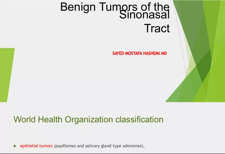
2. Benign Tumors of the Sinonasal Tract SAYED MOSTAFA HASHEMI MD
3. World Health Organization classification epithelial tumors (papillomas and salivary glandtype adenomas), soft tissue tumors (myxoma, leiomyoma, hemangioma,schwannoma, neurofibroma, and meningioma), tumors of bone and cartilage (giant cell lesion, giant cell tumor, chondroma, osteoma, chondroblastoma, chondromyxoid fibroma, osteochondroma, osteoid osteoma, osteoblastoma, ameloblastoma, and nasal chondromesenchymal hamartoma). Juvenile angiofibroma is traditionally included in the group of sinonasal tract tumors because of its common presentation as a nasal or nasopharyngeal mass
4. imaging techniques and the widespread application of endoscopic surgery have led to revived interest in the management of benign tumors of the Sinonasal tract
5. Clinical presentation Apart from osteomas, which are usually unexpectedly diagnosed on computed tomography (CT) of the brain or orbits or are associated with frontal headache in the absence of any other nasal complaint , The heralding symptom of most benign tumors of the sinonasal tract is unilateral nasal obstruction,which nowadays requires initial investigation with endoscopy of the nasal cavities after appropriate decongestion
6. Imaging studies common policy to obtain imaging studies, which can help to define the vascularization of the mass and its relationships with adjacent structures and to rule out a diagnosis of encephalomeningocele . In general, our preference is MRI because it can clearly differentiate tumor from retained secretions, allows higher contrast resolution, and may even suggest the nature of a soft tissue lesion. CT with contrast medium enhancement can be considered as a reasonable alternative
7. Diagnostic biopsy A biopsy is required whenever a diagnosis cannot be established by imaging studies but should be avoided if an angiofibroma is suspected
8. MANAGMENT Even though most benign tumors of the sinonasal tract can now be managed through an endoscopic approach, certain situations still require an external or a combined procedure. Need for an external or combined procedure may be clearly suggested by imaging studies , but there are cases in which a definitive decision can be made only during surgery. Consequently, the possibility of switching from an endoscopic to an external procedure must be discussed with the patient preoperatively
10. INVERTED PAPILLOMA Inverted papilloma included in the group of sinonasal papillomas, it is the second most common benign tumor of the sinonasal tract after osteoma, even though it represents the most common surgical indication for a benign sinonasal tumor. Incidence that ranges from 0.6 to 1.5 cases per 100,000 population annually. Men are more commonly affected than women, and the lesion is typically seen in the fifth and sixth decades of life.
11. site of origin Inverted papilloma most frequently arises from the lateral nasal wall in the fontanelle area. The maxillary sinus is the second most commonly affected site, and frontal and sphenoid sinuses are rarely involved primarily. Often the lesion extensively involves more than one sinus, making it impossible to assess the exact site of origin.
12. Diagnosis Endoscopy of the nose usually shows a pale, polypoid lesion with a papillary appearance that protrudes from the middle meatus this can easily suggest the diagnosis, which is sometimes made less obvious by the concomitant presence of inflammatory polyps.
13. common symptom Unilateral nasal obstruction with watery rhinorrhea is the most common symptom to prompt the patient to seek otolaryngologic consultation epiphora, proptosis, diplopia,and headache may be associated with an advanced lesion that involves the orbit or the skull base.
14. The association of inverted papilloma with scc The association of inverted papilloma with squamous cell carcinoma has been overemphasized, with reported frequencies as high as 56%. Recent data clearly show that the prevalence varies between 3.4% and 9.7% and that a synchronous occurrence is more common than a metachronous one.
15. The presence of human papillomavirus (HPV) Serotypes 16 and 18 have been specifically found to be associated with inverted papillomas that show histologic signs of malignant transformation.
16. Imaging studies Imaging studies are required to assess the extent and three dimensional configuration of the lesion and to disclose its relationship with surrounding structures (i.e., orbit, skull base, optic nerve, internal carotid artery) These goals are best achieved by MRI with gadolinium enhancement,.which also has the advantage over CT of better differentiating tumor from inflammatory mucosal changes.
17. CTS Numerous studies indicate that focal hyperostosis and osteitic changes seen on CT may be considered reliable predictors of tumor origin. These findings may also be identified by MRI
18. Surgical treatment Endoscopic surgery is a reliable alternative to traditional external techniques for the vast majority of lesions. However, an exclusively endoscopic approach may be contraindicated in the event of 1) massive involvement of the mucosa of the frontal sinus and/or of a supraorbital cell ; 2) transorbital extension, a very uncommon situation usually found in patients who have already undergone one or more surgical procedures; 3) concomitant presence of a malignancy that involves critical areas; 4)presence of significant scarring and anatomic distortion from previous surgery.
19. key point of endoscopic surgery To dissect the involved mucosa along the subperiosteal plane and to drill the underlying bone whenever required by imaging and/or intraoperative findings. The extent of the operation is dictated by the site of the lesion and the area of mucosa involved by the lesion.
20. Three basic types of endoscopic resections are Type I resection is indicated for inverted papillomas that involve the middle meatus, ethmoid, superior meatus, sphenoid sinus, or a combination of these structures; even lesions that protrude into the maxillary sinus without direct involvement of the mucosa are amenable to this approach.
21. Type II resection which corresponds to an endoscopic medial maxillectomy, is indicated for tumors that originate within the naso ethmoid complex and secondarily extend into the maxillary sinus or for primary maxillary lesions that do not involve the anterior and lateral walls of the sinus . The nasolacrimal duct can be included in the specimen to increase the exposure of the anterior part of the maxillary sinus.
22. Type III resection so known as the endonasal Denker operation , Entails removal of the medial portion of the anterior wall of the maxillary sinus to enable access to all the antrum walls. It is therefore recommended for the lamina papyracea.
23. Frontal sinus involvement Frontal sinus involvement can be observed in different scenarios that range from a limited lesion that grows marginally from the ethmoid into the frontal recess to very complex cases in which most or all of the sinus mucosa is diseased. Because the key point for ultimate success is removal of the lesion along the subperiosteal plane with the possibility to drill out the underlying bone, the surgical choice may involve a Draf IIB or Draf III endoscopic sinusotomy or a combination of an endoscopic approach with external sinusotomy through an osteoplastic flap,
24. FLOW UP The operation should end with the creation of a largely marsupialized cavity that will give wide access during follow-up for endoscopic inspection, which should be periodically performed to diagnose residual or recurrent lesions early. Postoperatively, MRI or CT is indicated only when a, the patient is symptomatic , or a residual or recurrent lesion has been histologically documented
25. outcomes The most recently published series clearly show that the recurrence rate is well below 10%. 17,26,27 Other than incomplete resection and high-stage disease , the risk of recurrence was recently found to be associated with cigarette smoking, 28 which also seems to favor malignant transformation. 29
26. JUVENILE ANGIOFIBROMA Juvenile angiofibroma is a benign lesion histologically characterized by vascular endotheliumlined spaces embedded in a fibrous stroma that typically affects young male adolescents. Immunohistochemical and electron microscopy studies suggest that the lesion could be considered a vascular malformation (or hamartoma) rather than a tumor.
27. JUVENILE ANGIOFIBROMA The lesion has a pathognomonic epicenter of origin at the level of the pterygopalatine fossa and subsequently grows through different pathways of spread that typically follow foramina and fissures of the skull base. The bone may be involved basically with two mechanisms: resorption by pressure coming from subperiosteal growth or invasion of the cancellous component, initially at the level of the root of the pterygoid process with subsequent expansion within the greater wing and erosion of the floor of the middle cranial fossa.
28. These observations led Schick and colleagues 36 to postulate that juvenile angiofibroma might develop from incomplete regression of a branchial artery, which arises in embryogenesis between days 22 and 24 and forms a temporary connection between the ventral aorta and dorsal aorta. This artery commonly regresses and forms a vascular plexus that either involutes or leaves remnants,which potentially leads to development of juvenile angiofibroma
30. Sign & symptom Unilateral nasal obstruction and epistaxis are the most common heralding symptoms of small to intermediate-size juvenile angiofibromas. In advanced lesions, swelling of the cheek,proptosis, or headache may be present, which indicates an involvement of the infratemporal fossa, the orbit, or the cranial fossa, respectively
31. The endoscopic finding The endoscopic finding of a smooth, hypervascularized lesion in a teenage boy that originates behind the middle turbinate, which is usually laterally displaced against the lateral wall strongly suggests a diagnosis of juvenile angiofibroma ;
32. Diagnosis of juvenile angiofibroma is based on imaging 1) The area of origin invariably located at the level of the pterygopalatine fossa, 2) its hypervascular appearance after contrast enhancement, 3) its pattern of growth. 39 On MRI, the presence on both T1- and T2-weighted sequences of several signal voids within the lesion, indicating major intralesional vessels,
33. Differential Diagnosis Sometimes differentiation of this lesion from lobular capillary hemangioma, hemangiopericytoma, and schwannoma can be difficult in view However, these other lesions do not commonly involve the pterygopalatine fossa and occur in a different age group.
34. Bleeding problem Although the currently available methods of embolization provide excellent devascularization of feeding vessels from the internal maxillary artery and its branches, as well as from the ascending pharyngeal artery, control of major vascularization from the internal carotid artery is challenging In case of encasement of the internal artery, which is indeed a very rare event, a balloon occlusion test and sacrifice of the internal carotid artery or, as a less invasive procedure, stenting of the intratemporal carotid artery may be considered.
35. embolization Not all authorities concur about routinely performing preoperative embolization. In fact, the modifications induced by this procedure at the tumor periphery have been shown to increase the likelihood of leaving residual tissue behind. 47
36. management of juvenile angiofibroma Surgery is considered the mainstay in the management of juvenile angiofibroma. Several approaches are currently available that range from microendoscopic techniques to midfacial degloving and infratemporal fossa resection
37. key steps The key steps to minimizing bleeding and achieving radical resection are the dissection of the lesion in the subperiosteal plane with the help of bipolar coagulation and an extensive drilling of the basisphenoid , where the tumor grows with digitations that are difficult to identify even under magnification. a dramatic reduction of recurrences with the systematic removal of the cancellous bone of the sphenoid involved by the lesion
38. alternatives The surgeon should also keep in mind that radiotherapy at a low dose (30 to 36 Gy) has been demonstrated to be effective in cases of advanced or recurrent lesions deemed not amenable to complete resection with acceptable morbidity. 57 The availability of new techniques such as Gamma Knife radiosurgery 58 and the Cyber knife system 59 might be alternatives with a low morbidity when used for lesions of limited size.
40. Postoperative surveillance Postoperative surveillance is based on periodic endoscopic and imaging examination, which should be performed for a minimum of 3 years. Because most of the residual lesions tend to grow submucosally and residual lesions are diagnosed within 1 year after surgery. , contrast-enhanced CT, or Preferably MRI,plays a key role in their early detection. Imaging studies performed in the immediate postoperative period would identify residual juvenile angiofibroma tissue more easily because of the absence of any inflammatory changes. most residual lesions are diagnosed within 1 year after surgery The actual frequency of residual lesions, although difficult to assess, ranges from 6% to 39% Treatment in the form of surgery or radiotherapy is scheduled only when there are clear signs of progressive increase in size. .
41. Postoperative surveillance Because the understanding of the biologic behavior of residual lesions is still limited, we commonly monitor residual lesions with MRI to assess their pattern of growth. Treatment in the form of surgery or radiotherapy is scheduled only when there are clear signs of progressive increase in size.
42. Osteoma Osteoma is a benign, slow-growing osteoblastic lesion and is the most common benign tumor of the sinonasal tract. It is found in 1% of patients undergoing plain sinus radiographs and in 3% of those undergoing CT for sinus symptoms. Osteomas are generally diagnosed between the second and fifth decades of life, with a slight male preponderance
43. Anatomic site The frontal sinus is the most frequently involved anatomic site (~80% of cases), followed by the ethmoid, the maxillary sinus, and, more rarely, the sphenoid sinus. Osteomas can be observed in conjunction with Gardner syndrome, a genetic disorder characterized by multiple polyps of the colon in association with osteomas of the skull and multiple soft tissue tumors.
44. main theories have been proposed 1) The embryologic theory proposes that osteoma develops at the junction between the embryonic cartilaginous ethmoid and the membranous frontal bone, 2)The traumatic theory correlates the development of osteoma with a previous trauma; 3) the infective theory is based on the belief that local inflammation may alter adjacent bone metabolism by activating osteogenesis. 4)slow-growing osseous hamartomas that arise in childhood.
45. Signs & Symptoms Most osteomas are asymptomatic and are occasionally diagnosed during radiographic examinations performed for unrelated reasons. osteoma may interfere with sinus drainage and can therefore cause sinusitis or even a mucocele with consequent frontal pain. If the lesion involves the outer table of the frontal bone, the patient may complain of aesthetic deformity , whereas tumors that grow toward the orbit can cause orbital or lacrimal symptoms (i.e., proptosis, diplopia, orbital pain, epiphora, decreased visual acuity, and even transient blindness). Osteomas that extend into the anterior cranial fossa carry the risk of cerebrospinal fluid leak, pneumocele, meningitis, andbrain abscess.
46. Endoscopic examination Endoscopic examination of the nasal cavity is usually normal, because the lesion is deeply located inside a paranasal cavity. Only in very rare instances, in which the osteoma grows toward the nasal cavity, can it be visualized as a firm mass covered by normal or atrophic mucosa
47. Imaging assessment Osteomas is currently based on CT .According to the amount of mineralized bone within the lesion, osteomas can exhibit a very high density that resembles cortical bone or a gradually decreasing density that displays a ground-glass pattern. Contrast enhancement is usually unnecessary to reach a diagnosis MRI may better delineate the relationship between the osteoma and adjacent soft tissues (i.e., dura, brain, orbital content).
48. Managment Because osteomas are slow-growing lesions, a general consensus in the literature advocates for a wait and see policy for any lesion that is asymptomatic, does not encroach upon critical structures such as the optic nerve (risk for intracranial complication), and does not extensively invade the anterior skull base or the orbit (risk for orbital complication). it is advisable to monitor the lesion with periodic cross sectional imaging examinations, which preferably include MRI.
49. surgical management Different external surgical approachesfrontoethmoidectomy through a Lynch-Howarth incision, midfacial degloving, lateralrhinotomy, Caldwell-Luc procedure, osteoplastic frontal sinusotomy dramatic change in the surgical management of these lesions, and most are now treated through a trans nasal approach
50. surgical management Cavitation is a surgical trick that helps resect even large osteomas through the nose; the core of the lesion is drilled with a cutting or diamond bur, leaving a very thin shell of bone that can be easily fractured and dissected from the adjacent tissues The possibility of using a device that induces ultrasound bone emulsification or performing piezosurgery has also been proposed. Cerebrospinal fluid leak can be expected during these maneuvers if the lesion is in contact with dura
51. postoperative surveillance Because osteoma recurrences are very rare, routine periodic postoperative surveillance by CT is not justified. We usually perform CT examination 1 year after surgery and, on the basis of its results, make a decision about further radiologic follow-up. The presence of a symptomatic stenotic frontal or maxillary sinusotomy is another situation that calls for CT examination
52. LOBULAR CAPILLARY HEMANGIOMA Lobular capillary hemangioma is a rapidly growing lesion characterized by a proliferation of capillaries arranged in lobules and separated by a loose connective tissue stroma, often infiltrated by inflammatory cells. The tumor has been commonly reported in the literature as pyogenic granuloma, which is currently considered inadequate in view of the fact that the lesion is neither the result of a bacterial infection nor a true granuloma.
53. lobular capillary hemangioma FIGURE 48-16. A lobular capillary hemangioma appears on a coronal contrast-enhanced magnetic resonance image as a large ethmoid lesion that abuts the orbit and encroaches on the floor of the anterior cranial fossa. The sharp borders of the intracranial part of the lesion and the subtle dural enhancement suggest extradural invasion ( arrows
54. etiopathogenesis The etiopathogenesis of lobular capillary hemangiomas is still obscure. Trauma, hormonal influences, viral oncogenes,
55. trauma The relevance of trauma such as habitual nose picking and nasal packing is supported by the fact that most lesions are located in the anterior half of the nasal cavity at the level of the Little area or on the head of the inferior and middle turbinates.
56. clinical presentation In its typical presentation, lobular capillary hemangioma appears at endoscopy as a red to purple mass, not larger than 1 cm, associated with epistaxis.75 However, in rarer instances, the lesion reaches a considerable size and fills the nasal cavity entirely, which leads to a complaint of unilateral nasal obstruction.
57. treatment Surgery is the treatment of choice for lobular capillary hemangioma, and radical resection can be performed through an endoscopic approach even in large lesions. 3 9
58. OSSIFYING FIBROMA AND FIBROUS DYSPLASIA fibrous dysplasia is a genetically based developmental anomaly of the bone-forming mesenchyme with a defect in osteoblastic differentiation and maturation that leads to replacement of normal bony tissue by fibrous tissue of variable cellularity and immature woven bone Ossifying fibroma Ossifying fibroma is a true benign neoplasm, main histologic finding are the absence of a capsule and the presence of more immature bone without osteoblastic activity in fibrous dysplasia
59. surgical treatment The objective of surgical treatment is different for the two lesions. Ossifying fibroma requires radical resection , in view of both the high rate of relapses which accounts for 44% of lesions localized in the ethmoids81and the aggressive behavior of recurring tumors, with local destruction and potential invasion of adjacent vital structures. Successful removal of ossifying fibroma via an endoscopic approach has been reported in the literature.
60. surgery For fibrous dysplasia surgery is intended to relieve symptoms , such as visual impairment as a result of compression of the optic nerve, or to correct aesthetic deformities. The type of resection, partial or radical, should be modulated based on the site of the lesion and consequently on the possibility of ensuing complications.86 Optic nerve decompression has been traditionally achieved through external transfacial,neurosurgical, or combined approaches, although most cases can be managed with endonasal endoscopic surgery
61. Alternative treatment Radiotherapy is contraindicated because of the risk of malignant transformation and the possible side effects on facial skeleton growth in young patients. Medical treatment of fibrous dysplasia based on the use of bisphosphonates (i.e., pamidronate), which inhibit osteoclastic activity, has been used with success in patients with extensive lesions associated with significant disfigurement and pain.92,93
62. SCHWANNOMA Schwannoma is a neurogenic tumor that arises from the Schwann cells of the sheath of myelinated nerves. This is a rare neoplasm that can be found in any part of the body; in 25% to 45% of cases, it is localized at the level of the head and neck. Only 4% of the lesions involve the sinonasal tract, where the ethmoid, maxillary sinus, nasal fossa, and sphenoid are involved (listed in order of decreasing frequency).
63. Sinonasal schwannomas Sinonasal schwannomas are considered to arise more frequently from the ophthalmic and maxillary divisions of the trigeminal nerves, but they can also originate from sympathetic fibers of the carotid plexus or from parasympathetic fibers of the pterygopalatine ganglion. Particularly in large lesions, identification of the nerve of origin at surgery is extremely difficult.
64. At endoscopy, the appearance of a sinonasal schwannoma is quite nonspecific; the presence of a network of capillaries on the surface can sometimes suggest a diagnosis of a hyper vascularlesion