Understanding Organ Rejection - The Science Behind It

In this informative article, we explore the reasons behind organ rejection. The mammalian immune system is a complex system that has evolved over millions of years in response to evolutionary stressors provided
- Uploaded on | 1 Views
-
 gayina
About Understanding Organ Rejection - The Science Behind It
PowerPoint presentation about 'Understanding Organ Rejection - The Science Behind It'. This presentation describes the topic on In this informative article, we explore the reasons behind organ rejection. The mammalian immune system is a complex system that has evolved over millions of years in response to evolutionary stressors provided. The key topics included in this slideshow are . Download this presentation absolutely free.
Presentation Transcript
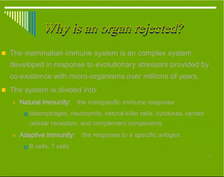
Slide11Why is an organ rejected? Why is an organ rejected? The mammalian immune system is an complex system developed in response to evolutionary stressors provided by co-existence with micro-organisms over millions of years. The system is divided into: Natural immunity: the nonspecific immune response Macrophages, neutrophils, natural killer cells, cytokines, certain cellular receptors, and complement components Adaptive immunity: the response to a specific antigen. B cells, T cells
Slide22Why is an organ rejected? Why is an organ rejected? In organ Tx, the principal target of the immune response to the graft are the Major Histocompatibility Complex (MHC) molecules expressed on the surface of donor cells (allo-MHC). This is a form of adaptive immunity.
Slide33 T cells recognize antigen in the form of peptide bound to MHC proteins. B cells have IG receptors that can recognize the antigenic portions of intact molecules.
Slide44T Cells T Cells T cells regulate the activities of B cells, T cells & other cells participating in immune responses. They provide help for Ab production by B cells, They are effectors of Ag-specific cell-mediated immunity (CMI). CMI is important in the elimination of Cells infected with intracellular pathogens (viruses, mycobacteria, some bacteria) Cells exhibiting aberrant differentiation (eg, neoplasms). Allogeneic cells (graft rejection).
Slide55Bretscher P, Cohn M: A theory of self-nonself discrimination. Science 169:1042-49, 1970
Slide66Activated T cells → Clonal expansion → Effector Mechanisms Activated T cells → Clonal expansion → Effector Mechanisms Mitogenic growth & differentiation factors. e.g IL-2. Mitogenic growth & differentiation factors. e.g IL-2.
Slide77Costimulation Allorocognition T cell activation Clonal Expansion of Alloreactive Cells Effector Mechanisms CD8 + T cells CD4 + T cells Apoptosis Induction Cell Lysis Macrophages B cells Allo-Ab Production DTH IL-2 Memor y T cells Th1 Th1 Th2 Th2 IL2 IFN IL2 IFN IL4, 5, 10, 13 IL4, 5, 10, 13
Slide88
Slide99MHC class I MHC class I
Slide1010MHC class I molecules are found on almost all cell types except for RBCs MHC class I molecules are found on almost all cell types except for RBCs Intra-cytoplasmic peptides are presented together with class I MHC on the cell surface to CD8 T cells . CD8 positive T cells induce cell lysis (by inducing apoptosis or active killing of cells)
Slide1111MHC class II MHC class II
Slide1212Class II MHC is expressed only on interstitial dendritic cells, macrophages, & B cells and activated endothelial cells. Class II MHC is expressed only on interstitial dendritic cells, macrophages, & B cells and activated endothelial cells. MHC II molecules bind peptides derived from extracellular (exogenous) proteins. Exogenous proteins that have been endocytosed and fragmented are transferred to the cell surface and presented to CD4 T cells together with class II MHC molecules.
Slide1313High levels of class II MHC are expressed on APCs High levels of class II MHC are expressed on APCs 1. Dendritic cells : are the major "professional" APCs. 2. Monocyte/Macrophages: play a partial role in Ag presentation 3. B cells : express MHC II, required to receive T cell help. Although they do not activate resting naive T cells, they may stimulate memory T cells. 4. Vascular endothelial cells.
Slide1414
Slide1515Other antigens mediating rejection Minor Histocompatibility Ags Other antigens mediating rejection Minor Histocompatibility Ags NIH GUIDE, Volume 26, Number 24, July 25, 1997 NIH GUIDE, Volume 26, Number 24, July 25, 1997 These antigens appear to be MHC class I bound small peptides presented to - -T cells in an MHC-restricted manner. Thus, mHAgs are recognized with the same degree of specificity as the major histocompatibility antigens Most mHAgs result from nonsynonymous Single Nucleotide Polymorphisms (SNP), accounting for around 90% of sequence differences (Collins et al, 1998). They elicit most commonly, MHC I restricted responses. They donnot activate Ab production, though they need CD4+ activation for CD8+ help ABO Antigens ABO Antigens Monocyte endothelial Antigens: Monocyte endothelial Antigens: Rarely cause hypercute rejection despite ABO compability Rarely cause hypercute rejection despite ABO compability
Slide1616Pathways of Allorcognition Pathways of Allorcognition Pathways of Allorcognition Pathways of Allorcognition
Slide1717Game DS, Lechler RI, Transplant Immunology, 2002 ,10(2) pp. 101-108(8)
Slide1818Normal Normal pathway pathway Early Acute rejection Early Acute rejection
Slide1919Ben M. Illigens et al, Human Immunology:2002, 63, 912–925
Slide2020Signal 1 Signal 1 Signal 2 Signal 2 Bretscher P, Cohn M: A theory of self-nonself discrimination. Science 169:1042-49, 1970 T cell activation and function T cell activation and function TK p651ck TK p651ck Ph. of IC proteins Ph. of IC proteins Activation of calcineurin, NFAT, NF-kB Activation of calcineurin, NFAT, NF-kB
Slide2121ZAP- 70 Fyn Lc k Cellular and Molecular Immunology (5th Ed.) Abbas AK, & Lichtman, Editor: Saunders, Philadelphia, 2005
Slide2222Resting T cells express CD28, but resting antigen-presenting cells (APCs) do not express B7 molecules. Within six hours after activation, B7-2 is expressed by antigen-presenting cells and is available to bind to CD28, transmitting a costimulatory signal to the T cell. By 48 to 72 hours after activation, antigen-presenting cells also express B7-1, whereas T cells express the CTLA-4 inhibitory receptor. Both B7-1 and B7-2 can bind to either CD28 or CTLA-4, providing continued costimulation or a new inhibitory signal, respectively. Because CTLA-4 binds B7 molecules with a higher affinity than does CD28, its inhibitory interaction eventually predominates, leading to the termination of the immune response. The fusion protein CTLA- 4–Ig can compete with CD28 and CTLA-4 for B7 binding, thus preventing costimulatory interactions. Sayegh MH, Turka LA NEJM, 1998, 338: 1813-21 6 hrs IL-2 & other cytokines Anergy or apoptos is
Slide2323Resting antigen-presenting cells (APCs), which include B cells, macrophages, and dendritic cells, express CD40. When activated, T cells express CD40 ligand (CD40L). Interactions between CD40 ligand and CD40 are important in providing B-cell help to prevent apoptosis and induce immunoglobulin production and isotype switching. Activation of CD40 on APCs provides a signal for the induction of B7 molecules, particularly B7-2. CD40 ligand may act in T-cell costimulation by directly providing costimulation, by inducing B7, or by inducing other costimulatory ligands. Sayegh MH, Turka LA NEJM, 1998, 338: 1813-21
Slide2424Accessory molecules of T cells Accessory molecules of T cells CD 28- B7: Production of IL-2 and other cytokines CTLA4- B7: Inhibition of T cell stimulation [Abatacept (CTLA4-Ig)] LFA-1- ICAM-1 (2, 3): Adhesion of T cells to APCs, endothelial cells and extracellular matrix proteins and adhesion-dependent lymphocyte functions L-Selectin-CH ligands on HEV: Regulate migration of leukocytes to various tissues CD44-matrix proteins: Retention of T cells to endothelium at sites of inflammation CD40L- CD40: important signals for Ab production Activation of macrophages to destroy phagocytosed microbes B7 expression on all APCs (role in co-stimulation) Production of adhesion molecules & inflammatory cytokines by APCs → T- cell activation Fas ligand(D95)- Fas: T cell apoptosis and elimination
Slide2525Accessory molecules of T cells Accessory molecules of T cells CD 28- B7: Production of IL-2 and other cytokines CD 28- B7: Production of IL-2 and other cytokines CTLA4- B7: Inhibition of T cell stimulation [Abatacept (CTLA4-Ig)] LFA-1- ICAM-1 (2, 3): Adhesion of T cells to APCs, endothelial cells and extracellular matrix proteins and adhesion-dependent lymphocyte functions L-Selectin-CH ligands on HEV: Regulate migration of leukocytes to various tissues CD44-matrix proteins: Retention of T cells to endothelium at sites of inflammation CD40L- CD40: important signals for Ab production Activation of macrophages to destroy phagocytosed microbes B7 expression on all APCs (role in co-stimulation) Production of adhesion molecules & inflammatory cytokines by APCs → T- cell activation Fas ligand(D95)- Fas: T cell apoptosis and elimination
Slide2626Accessory molecules of T cells Accessory molecules of T cells CD 28- B7: Production of IL-2 and other cytokines CTLA4- B7: Inhibition of T cell stimulation [Abatacept (CTLA4-Ig)] CTLA4- B7: Inhibition of T cell stimulation [Abatacept (CTLA4-Ig)] LFA-1- ICAM-1 (2, 3): Adhesion of T cells to APCs, endothelial cells and extracellular matrix proteins and adhesion-dependent lymphocyte functions L-Selectin-CH ligands on HEV: Regulate migration of leukocytes to various tissues CD44-matrix proteins: Retention of T cells to endothelium at sites of inflammation CD40L- CD40: important signals for Ab production Activation of macrophages to destroy phagocytosed microbes B7 expression on all APCs (role in co-stimulation) Production of adhesion molecules & inflammatory cytokines by APCs → T- cell activation Fas ligand(D95)- Fas: T cell apoptosis and elimination
Slide2727Accessory molecules of T cells Accessory molecules of T cells CD 28- B7: Production of IL-2 and other cytokines CTLA4- B7: Inhibition of T cell stimulation [Abatacept (CTLA4-Ig)] LFA-1- ICAM-1 (2, 3): Adhesion of T cells to APCs, endothelial cells and extracellular matrix proteins & adhesion-dependent lymphocyte functions LFA-1- ICAM-1 (2, 3): Adhesion of T cells to APCs, endothelial cells and extracellular matrix proteins & adhesion-dependent lymphocyte functions L-Selectin-CH ligands on HEV: Regulate migration of leukocytes to various tissues CD44-matrix proteins: Retention of T cells to endothelium at sites of inflammation CD40L- CD40: important signals for Ab production Activation of macrophages to destroy phagocytosed microbes B7 expression on all APCs (role in co-stimulation) Production of adhesion molecules & inflammatory cytokines by APCs → T- cell activation Fas ligand(D95)- Fas: T cell apoptosis and elimination
Slide2828Accessory molecules of T cells Accessory molecules of T cells CD 28- B7: Production of IL-2 and other cytokines CTLA4- B7: Inhibition of T cell stimulation [Abatacept (CTLA4-Ig)] LFA-1- ICAM-1 (2, 3): Adhesion of T cells to APCs, endothelial cells and extracellular matrix proteins and adhesion-dependent lymphocyte functions L-Selectin-CH ligands on HEV: Regulate migration of leukocytes to various tissues L-Selectin-CH ligands on HEV: Regulate migration of leukocytes to various tissues CD44-matrix proteins: Retention of T cells to endothelium at sites of inflammation CD40L- CD40: important signals for Ab production Activation of macrophages to destroy phagocytosed microbes B7 expression on all APCs (role in co-stimulation) Production of adhesion molecules & inflammatory cytokines by APCs → T- cell activation Fas ligand(D95)- Fas: T cell apoptosis and elimination
Slide2929Accessory molecules of T cells Accessory molecules of T cells CD 28- B7: Production of IL-2 and other cytokines CTLA4- B7: Inhibition of T cell stimulation [Abatacept (CTLA4-Ig)] LFA-1- ICAM-1 (2, 3): Adhesion of T cells to APCs, endothelial cells and extracellular matrix proteins and adhesion-dependent lymphocyte functions L-Selectin-CH ligands on HEV: Regulate migration of leukocytes to various tissues CD44-matrix proteins: Retention of T cells to endothelium at sites of inflammation CD44-matrix proteins: Retention of T cells to endothelium at sites of inflammation CD40L- CD40: important signals for Ab production Activation of macrophages to destroy phagocytosed microbes B7 expression on all APCs (role in co-stimulation) Production of adhesion molecules & inflammatory cytokines by APCs → T- cell activation Fas ligand(D95)- Fas: T cell apoptosis and elimination
Slide3030Accessory molecules of T cells Accessory molecules of T cells CD 28- B7: Production of IL-2 and other cytokines CTLA4- B7: Inhibition of T cell stimulation [Abatacept (CTLA4-Ig)] LFA-1- ICAM-1 (2, 3): Adhesion of T cells to APCs, endothelial cells and extracellular matrix proteins and adhesion-dependent lymphocyte functions L-Selectin-CH ligands on HEV: Regulate migration of leukocytes to various tissues CD44-matrix proteins: Retention of T cells to endothelium at sites of inflammation CD40L- CD40: CD40L- CD40: important signals for Ab production important signals for Ab production Activation of macrophages to destroy phagocytosed microbes Activation of macrophages to destroy phagocytosed microbes B7 expression on all APCs (role in co-stimulation) B7 expression on all APCs (role in co-stimulation) Production of adhesion molecules & inflammatory cytokines by APCs → T- cell activation Production of adhesion molecules & inflammatory cytokines by APCs → T- cell activation Fas ligand(D95)- Fas: T cell apoptosis and elimination
Slide3131Accessory molecules of T cells Accessory molecules of T cells CD 28- B7: Production of IL-2 and other cytokines CTLA4- B7: Inhibition of T cell stimulation [Abatacept (CTLA4-Ig)] LFA-1- ICAM-1 (2, 3): Adhesion of T cells to APCs, endothelial cells and extracellular matrix proteins and adhesion-dependent lymphocyte functions L-Selectin-CH ligands on HEV: Regulate migration of leukocytes to various tissues CD44-matrix proteins: Retention of T cells to endothelium at sites of inflammation CD40L- CD40: important signals for Ab production Activation of macrophages to destroy phagocytosed microbes B7 expression on all APCs (role in co-stimulation) Production of adhesion molecules & inflammatory cytokines by APCs → T- cell activation Fas ligand (D95)- Fas: T cell apoptosis and elimination Fas ligand (D95)- Fas: T cell apoptosis and elimination
Slide3232Principle accessory molecules of T Lymphocytes Principle accessory molecules of T Lymphocytes Name Gene family Cellular expression Ligand Role in T cell activation Adhesion Signaling CD4 Ig Class II T cells Class II MHC ± + CD8 Ig Class I T cells Class I MHC ± + CD28 Ig >90% CD4+ T cells, ~50% D8+ T cells B7-1 (CD 80) B7-2 (CD 86) - + CTLA-4 (CD 152) Ig Activated T cells B7-1 B7-2 - + CD45R Leukocytes Unknown - + CD2 (LFA-2) Ig >90% T cells (human), NK cells LFA-3 + + LFA-1 Integrin Leukocytes, platelets ICAM-1 ICAM-2 ICAm-3 + ± L-selectin Selectin Leukocytes Carbohydrate ligands on HEV + - CD44 Lymphocytes, granulocytes Matrix proteins + ± Cellular and Molecular Immunology (5th Ed.) Abbas AK, & Lichtman, Editor: Saunders, Philadelphia, 2005.
Slide3333Salama AD, Sayegh MH. Am J Transplant 2003; 3: 509–511 (B7-1) (B7-2) Alternative T-Cell Costimulatory Pathways in Alternative T-Cell Costimulatory Pathways in Transplant Rejection and Tolerance Induction Transplant Rejection and Tolerance Induction
Slide3434
Slide3535Intracellular events Intracellular events
Slide3636
Slide3737Immunoreceptor Tyrosine Based Activating Motifs ζζ Isakov N - J Leukoc Biol 1997 Jan;61(1):6-16. CD4, CD8 NF- қ B Lck Fyn ZAP-70 CD3 LAT SLP-76 Grb2 Shc Sos Ras MAP-K AP-1 Jun Fos LAT SLP-76 Vav p110 p85 Rho Family GTPase Regulatoroy Function for Cytoskelton PI-3 K LAT SLP-76 PLC- γ 1 IP-3 DAG PLC- γ 1 Ca 2 + calcineurin NF-AT p Adaptor proteins: Protein-Protein interaction • SLP-76 • Lat • GRb • VAV dephosphorylate TCR complex Itams
Slide38381, Cyclosporine 2, Tacrolimus (FK506) 3, Rapamycin 4, Corticosteroid 1 2 3 4
Slide3939L spleen, lymph nodes, Peyer's patches T Scan information about new immunological events peripheral tissues T T T T T T T D Toll-like receptor
Slide4040Tolerance Tolerance Central (Negative Selection): Central (Negative Selection): Immature lymphocytes in the generative lymphoid organs (thymus for T cells and bone marrow for B cells) encounter only self Ags in high concentration . These lymphyctes are killed or deleted. Some of the T cells may develop into regulatory cells → Inhibition of immune response Peripheral: Peripheral: Important for maintaining unresponsiveness to those self Ags that are expressed in peripheral lymphoid tissues (spleen, lymph nodes, mucosal lymphoid)
Slide4141Mechanisms of Tolerance Mechanisms of Tolerance 1. Deletion: Apoptotic cell death 1. Deletion: Apoptotic cell death Activation induced cell death: Fas-FasL interaction Binding if FAAD Activation of caspase 8 Activation of effector caspases Apoptosis Passive cell death (death by neglect) Loss of survival stimuli e.g. growth factors or costimulators Increase mitochondrial permeability Release of cytochrome c and then Apaf Activation of caspase 9 Activation of effector caspases Apoptosis
Slide4242Mechanisms of Tolerance… Mechanisms of Tolerance… 2. Anergy: Functional inactivation without cell death 2. Anergy: Functional inactivation without cell death a. Ag presentation by APCs without costimulation (Signal 2 or B7-CD28) b. Engagement of inhibitory receptors (CTLA-4 instead of CD28) CTLA-4 Ig: Abatacept, LEA29Y (belatacept) Antibodies against CD40L
Slide4343Mechanisms of Tolerance… Mechanisms of Tolerance… 3. Supression: by regulatory lymphocytes 3. Supression: by regulatory lymphocytes These are CD4+ T cells expressing high levels of IL-2-R (CD25) but not other markers of activation Inhibition of these cells may cause autoimmune disease 4. Clonal ignorance: 4. Clonal ignorance: Self Ags may be ignored by the immune system, i.e. lymphocytes encounter the Ag but fail to respond
Slide4444Auchincloss H, Am J Transplant 2001;1(6-12) Auchincloss H, Am J Transplant 2001;1(6-12) Auchincloss H, Am J Transplant 2001;1(6-12) Auchincloss H, Am J Transplant 2001;1(6-12) The Thymic Deletional Mechanism The Thymic Deletional Mechanism Exposure to donor antigens during fetal development achieved the same thymic deletional tolerance that occurs for many self antigens. Engraftment of donor stem cells Mixed bone marrow chimeras Thymic deletion of T cells reactive to donor APCs Re-poplulation of a new T cell repertoire
Slide4545Auchincloss H, Am J Transplant 2001;1(6-12) Auchincloss H, Am J Transplant 2001;1(6-12) Auchincloss H, Am J Transplant 2001;1(6-12) Auchincloss H, Am J Transplant 2001;1(6-12) Peripheral Deletional Mechanisms Peripheral Deletional Mechanisms Removal donor-specific T cells after they have matured. Mechanisms: Veto phenomenon: involves the ability of a population of CD8 T cells to destroy attacking T cells that would otherwise kill them, via attachment to their MHC I molecules. Cell death mediated through Fas Ligand, Other forms of activation-inducedcell death (AICD) Withdrawal of growth factors such as IL-2. Peripheral Nondepleting Mechanisms Peripheral Nondepleting Mechanisms An active regulation of T cell responses by other (suppressor) T cells Change in the character of the donor specific response
Slide4646 Although multiple mechanisms of tolerance induction may be combined, there are two hallmarks of a tolerance induction strategy that will ultimately be successful in clinical practice: It will include the use of donor bone marrow (or stem cells) somewhere in the protocol, It will eventually lead to easily measurable macrochimerism. Auchincloss H, Am J Transplant 2001;1(6-12) Auchincloss H, Am J Transplant 2001;1(6-12) Auchincloss H, Am J Transplant 2001;1(6-12) Auchincloss H, Am J Transplant 2001;1(6-12)
Slide4747Graft rejection is usually initiated by CD4 helper T cells (TH) that bind peptides in complexes with MHC class II molecules on antigen- presenting cells. In the direct pathway of recognition, an MHC molecule on a foreign (allogeneic) cell, such as an antigen-presenting cell (allogeneic APC), binds to the helper T cell. In the indirect pathway, the foreign MHC molecule is processed into peptides that are presented to the helper T cell by one of the body's own antigen-presenting cells (self APC). In either case, activated CD4 helper T cells proliferate and secrete a variety of cytokines that serve as growth and activation factors for CD8 cytotoxic T cells (TC), B cells, and macrophages, which cause destruction of the graft by direct lysis of target cells, antibody production, and delayed-type hypersensitivity mechanisms, respectively. Sayegh MH, Turka LA NEJM, 1998, 338: 1813-21