Understanding the Sensory System: Receptors and Stimuli

This article provides an introduction to the sensory system and explains the different receptors that detect environmental changes and stimuli. It covers chemoreceptors, pain receptors, thermoreceptors, mechanoreceptors, and photoreceptors.
- Uploaded on | 0 Views
-
 tonypagac
About Understanding the Sensory System: Receptors and Stimuli
PowerPoint presentation about 'Understanding the Sensory System: Receptors and Stimuli'. This presentation describes the topic on This article provides an introduction to the sensory system and explains the different receptors that detect environmental changes and stimuli. It covers chemoreceptors, pain receptors, thermoreceptors, mechanoreceptors, and photoreceptors.. The key topics included in this slideshow are sensory system, receptors, stimuli, chemoreceptors, pain receptors, thermoreceptors, mechanoreceptors, photoreceptors,. Download this presentation absolutely free.
Presentation Transcript
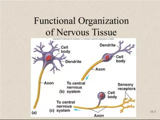
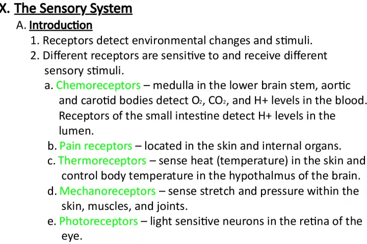
2. X. The Sensory System A. Introduction 1. Receptors detect environmental changes and stimuli. 2. Different receptors are sensitive to and receive different sensory stimuli. a. Chemoreceptors medulla in the lower brain stem, aortic and carotid bodies detect O 2 , CO 2 , and H+ levels in the blood. Receptors of the small intestine detect H+ levels in the lumen. b. Pain receptors located in the skin and internal organs. c. Thermoreceptors sense heat (temperature) in the skin and control body temperature in the hypothalmus of the brain. d. Mechanoreceptors sense stretch and pressure within the skin, muscles, and joints. e. Photoreceptors light sensitive neurons in the retina of the eye.
5. 3. Sensation a. Sensations are feelings resulting from sensory stimulation. b. A particular part of the sensory cortex always interprets impulses reaching it in the same way. c. The brain projects a sensation back to the region of stimulation. 4. Sensory adaptations are adjustments made by sensory receptors to continuous stimulation in which impulses are triggered at slower and slower rates. B. Pain receptors 1. Pain receptors are free nerve endings stimulated by tissue damage. 2. The only receptors in visceral organs that provide sensation are pain receptors , ( visceral pain ). 3. The sensations produced from visceral receptors are likely to feel as if they are coming from some other part or location. This is called referred pain.
6. 4. Somatic pain originates in the skin, skeletal muscles, joints and tendons. 5. Stimulated nerve fibers from amputated limbs can tend to result in phantom pain . 6. Pain experience has two components: pain stimulus and reaction to pain stimulus which determines the amount of suffering .
7. C. Sense of Smell (Olfaction) 1. Olfactory organs a. The olfactory organs consist of receptors and supporting cells in the nasal cavity . b. Olfactory receptors are neurons with cilia that are sensitive to gaseous/dissolved chemicals . c. Nerve impulses travel from the olfactory receptors through the olfactory nerves , olfactory bulbs , and olfactory tracts to interpreting centers in the olfactory portion of the cerebral cortex . You perceive odors (smells) in your brain! 2. Olfactory stimulation a. Olfactory impulses may result when various gaseous molecules combine with specific binding sites on the cilia of the receptor cells. b. Olfactory receptors adapt rapidly.
9. D. Sense of Taste 1. Taste receptors a. Taste buds consist of receptor cells and supporting cells located in papillae on the tongue . b. Taste cells have taste hairs that are sensitive to particular chemicals dissolved in water . c. Taste hair surfaces seem to have receptor sites to which chemicals combine. 2. Taste sensation ( gustation ) a. The five primary taste sensations are: sweet , sour , salty , bitter , and umami . b. Various taste sensations result from the stimulation of one or more sets of taste receptors. c. Much of taste also involves the sense of smell.
11. E. Sense of Hearing 1. The external ear composed of three parts collects sound waves created by vibrating objects. a. The visible ear that collects and directs sound is the auricle or pinna . b. The external auditory meatus directs sound into the skull. c. The tympanic membrane vibrates to begin the conversion of sound energy to mechanical energy. 2. Middle ear (air filled space) a. The three auditory ossicles , ( malleus , incus , & stapes ) of the middle ear conduct and multiply the energy of vibration to the oval window of the inner ear. b. Eustachian tubes connect the middle ear to the throat and function to help maintain equal air pressure on both sides of the eardrums so they are free to vibrate in response to sound.
15. 3. Inner ear a. The inner ear consists of a complex system of interconnected tubes and chambers, the osseous ( bony ) filled with perilymph, and membranous (membrane) labyrinths filled with endolymph. b. The organ of Corti contains the hearing receptors ( hair cells ) that are stimulated by vibrations in the fluids of the inner ear. c. Different frequencies of vibrations are thought to stimulate different receptor hair cells. 4. Steps in hearing : a. Sound waves collected by auricle ( pinna ) b. Waves pass through External Auditory Meatus c. Tympanic Membrane vibrates d. Malleus connected on the medial side of Tympanic Membrane moves. e. Malleus moves the incus f. Incus moves the stapes g. Stapes pushes on the oval window . Movement of stapes is 20 times greater than that of the Tympanic Membrane
16. h. Perilymph in Scala Vestibuli moves i. Perilymph wave energy causes the Basilar Membrane in the cochlear duct to move up and down. j. Organ of Corti hair cells within the cochlear duct (containing endolymph ) move up and down k. Hair cells touch the Tectorial Membrane l. Hairs bend and send nerve impulses down the cochlear branch of the Vestibulocochlear Nerve to the temporal lobe of the cerebrum. m. Different frequencies cause different regions of the Basilar Membrane to vibrate causing different regions of Organ of Corti hair cells to be stimulated = perception of different frequencies. o. Movement of perilymph in the Scala Vestibuli goes to the end of the cochlea and moves the perilymph in the Scala Tympani which pushes on the Round Window so the wave energy is lost, hearing stops.
18. F. Sense of Equilibrium 1. Static equilibrium is concerned with maintaining the stability of the head and body when these parts are motionless .
20. 2 . Dynamic equilibrium is concerned with balancing the head and body when they are moved or rotated suddenly . Sensory organs of dynamic equilibrium are located in the Semicircular canals .
22. 3. Other parts that help with the maintenance of equilibrium include the eyes and mechanoreceptors associated with certain joints called proprioceptors . G. Sense of Sight 1. Visual accessory organs include the eyelids, lacrimal apparatus ( lacrimal gland produces tears , Nasolacrimal duct drains the eye to the nasal cavity), and extrinsic muscles to move the eye.
24. 2. Structure of the Eye a. The wall of the eye has an outer, middle, and inner layer that functions as follows: 1.) The outer white layer ( sclera ) is fibrous, protective , and shapes the eye . Its transparent anterior portion ( cornea ) protects and refracts (bends) light entering the eye. Astigmatism is blurry vision in parts of the visual field caused by unequal curvatures waviness in the cornea. 2.) The middle layer ( choroid ) is the vascular layer and contains a brown pigment that helps to absorb light to avoid visual confusion. 3.) the inner layer ( retina ) contains the visual receptor cells b. The lens is a transparent, elastic structure whose shape is controlled by the action of the ciliary muscles that are part of the ciliary body . The lens changes shape to refract light. c. The iris is a muscular diaphragm that controls the amount of light entering the eye. The pupil is the hole in the middle of the iris.
25. d . S p a c e s w i t h i n t h e e y e a r e f i l l e d w i t h f l u i d s t h a t h e l p t o m a i n t a i n t h e s h a p e o f t h e e y e . 1 . ) A n t e r i o r c a v i t y ( i n f r o n t o f t h e l e n s ) i s f i l l e d w i t h a q u e o u s h u m o r 2 . ) P o s t e r i o r c a v i t y ( l a r g e r a n d b e h i n d t h e l e n s ) i s f i l l e d b y v i t r e o u s h u m o r . 3 . R e f r a c t i o n o f l i g h t a . L i g h t w a v e s a r e r e f r a c t e d p r i m a r i l y b y t h e c o r n e a a n d l e n s . b . T h e l e n s m u s t b e t h i c k e n e d ( a c c o m m o d a t i o n ) t o f o c u s o n o b j e c t s c l o s e r t h a n 2 0 f e e t a w a y b y u s e o f t h e c i l i a r y m u s c l e s . 4 . V i s u a l r e c e p t o r s a . T h e v i s u a l r e c e p t o r s a r e c a l l e d r o d s a n d c o n e s 1 . ) R o d s ( m o r e s e n s i t i v e t o l o w l i g h t ) p r o v i d e b l a c k & w h i t e v i s i o n t h a t i s p o o r i n d e t a i l 2 . ) C o n e s r e q u i r e h i g h e r i n t e n s i t y l i g h t t o p r o v i d e h i g h l y d e t a i l e d c o l o r v i s i o n .
28. b. Visual pigments 1.) A light-sensitive pigment in rods decomposes in the presence of light to trigger nerve impulses that our visual cortex perceives as vision 2.) Color vision seems to be related to the presence of three sets of cones ( blue , green , red ) containing different light-sensitive pigments. Cones are most highly concentrated at the Macula Lutea of the retina. This is the area of most acute color vision and light hits it when you look directly at objects. 5. Stereoscopic vision a. Stereoscopic vision involves the perception of distance and depth ( depth perception ) b. Stereoscopic vision occurs because of the formation of two slightly different retinal images that the brain superimposes and interprets as one image in three dimensions
29. 6. Visual nerve pathways a. Nerve fibers from the retina form the optic nerves . Nerve fibers exit the back of the eye at the optic disc causing the blind spot . Because nerve fibers fill this area, no cones or rods are present. b. Some nerve fibers from each eye cross over in the optic chiasm . This ensures that all areas of the visual field perceived by both eyes are processed by one side of the brain into a coherent image.
30. 7. Shape of the eyeball a. If elongate ( too long ): 1.) The focus point of the image is in front of the retina , the image that hits the retina is out of focus. 2.) This is Myopia short sighted vision you cant see far away so you are described as being nearsighted . b. If eyeball is ( too short ): 1.) The focus point of the image occurs past ( behind ) the retina so the image that hits the retina is out of focus. 2.) This is hyperopia farsightedness You cant see close objects!
31. 8. Aging a. As you get older the lens gets harder and will not thicken as or accommodate as much for close vision. This is usually corrected by glasses or contact lenses.