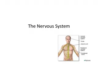
Functional Properties of Neurons: Irritability, Conductivity, and Starting a Nerve Impulse

This article explores the functional properties of neurons, including their ability to respond to stimuli (irritability) and transmit impulses (
- Uploaded on | 30 Views
-
 almapedersen
About Functional Properties of Neurons: Irritability, Conductivity, and Starting a Nerve Impulse
PowerPoint presentation about 'Functional Properties of Neurons: Irritability, Conductivity, and Starting a Nerve Impulse'. This presentation describes the topic on This article explores the functional properties of neurons, including their ability to respond to stimuli (irritability) and transmit impulses (. The key topics included in this slideshow are . Download this presentation absolutely free.
Presentation Transcript
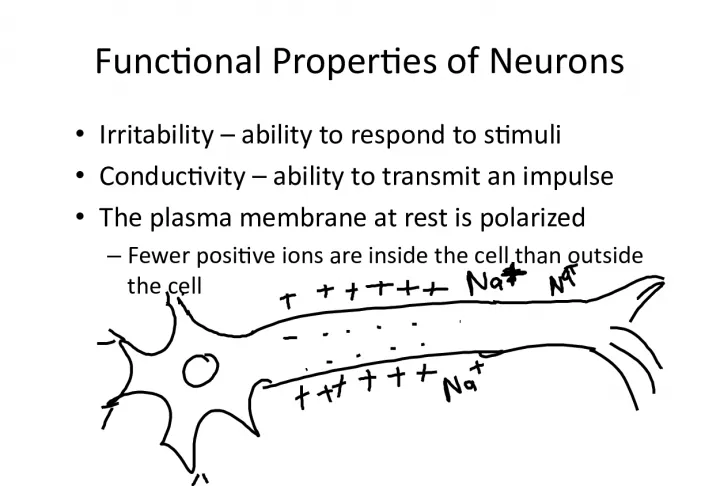
Slide1Functional Properties of Neurons• Irritability – ability to respond to stimuli • Conductivity – ability to transmit an impulse • The plasma membrane at rest is polarized – Fewer positive ions are inside the cell than outside the cell
Slide2Starting a Nerve Impulse• Stimulus : something that causes a neuron to start an impulse • Depolarization – a stimulus depolarizes the neuron ’ s membrane – A depolarized membrane allows sodium (Na + ) to flow inside the membrane • The exchange of ions initiates an action potential in the neuron Figure 7.9a–c
Slide3The Action Potential If the action potential (nerve impulse) starts, it is propagated over the entire axon Potassium ions rush out of the neuron after sodium ions rush in, which repolarizes the membrane The sodium-potassium pump restores the original configuration This action requires ATP
Slide4Nerve Impulse Propagation The impulse continues to move toward the cell body Impulses travel faster when fibers have a myelin sheath made of Schwann cells Figure 7.9d–f nerve impulse animation
Slide5Continuation of the Nerve Impulsebetween Neurons • Impulses are able to cross the synapse (space between neurons) to another neuron – Neurotransmitter (chemical messenger) is released from a nerve ’ s axon terminal – The dendrite of the next neuron has receptors that are stimulated by the neurotransmitter – An action potential is started in the dendrite
Slide6How NeuronsCommunicate at Synapses
Slide7Reflex• Reflex – rapid, predictable, and involuntary response to stimuli Figure 7.11a
Slide8Types of Reflexes and Regulation• Somatic reflexes – Activation of skeletal muscles • Autonomic reflexes – Smooth muscle regulation – Heart and blood pressure regulation – Regulation of glands – Digestive system regulation
Slide9The Reflex Arc• Reflex arc – direct route from a sensory neuron, to an interneuron, to a motor neuron, to an effector
Slide10Elements of Reflex ArcFigure 7.11b–c 1. Sensory receptor – receives the stimulus 2. Sensory neuron – carries impulse to association neuron/interneuron in CNS 3. Integration center – made of interneurons in CNS (either brain or spinal cord) 4. Motor neuron – carries impulse to effector from CNS 5. Effector organ – muscle or gland Reflex arc
Slide11CNS: Regions of the Brain• Cerebrum/Cerebral hemispheres • Diencephalon • Brain stem • Cerebellum Figure 7.12b
Slide12Cerebral Hemispheres (Cerebrum)• Paired (left and right) superior parts of the brain • Include more than half of the brain mass Figure 7.13a
Slide13Cerebral Hemispheres (Cerebrum)• The surface is made of ridges (gyri) and grooves (sulci) Figure 7.13a
Slide14Lobes of the Cerebrum• Fissures (deep grooves) divide the cerebrum into lobes • Surface lobes of the cerebrum – Frontal lobe – motor control, motor speech, language comprehension, emotions – Parietal lobe – tasting, speech comprehension – Temporal lobe – hearing, smell – Occipital lobe - sight
Slide15Lobes of the CerebrumFigure 7.15a
Slide16Specialized Areas of the CerebrumFigure 7.13c • Somatic sensory area – receives impulses from the body’s sensory receptors (parietal lobe) • Primary motor area – sends impulses to skeletal muscles (frontal lobe) • Broca’s area – involved in our ability to speak (frontal lobe)
Slide17Sensory and Motor Areas of theCerebral Cortex Figure 7.14 How do you think scientists developed this graphic? What tests were done ?
Slide18Sensory Areas of the Cerebral CortexFigure 7.14
Slide19Layers of the Cerebrum• Gray matter – Outer layer – Composed mostly of neuron cell bodies Figure 7.13a
Slide20Layers of the Cerebrum• White matter – Fiber tracts inside the gray matter – Example: corpus callosum connects hemispheres Figure 7.13a
Slide21Diencephalon• Sits on top of the brain stem • Enclosed by the cerebral hemispheres • Made of three parts – Thalamus – Hypothalamus – Epithalamus
Slide22Thalamus• The relay station for sensory impulses • Transfers impulses to the correct part of the cortex for localization and interpretation
Slide23Hypothalamus• Under the thalamus • Important autonomic nervous system center – Helps regulate body temperature – Controls water balance – Regulates metabolism
Slide24Hypothalamus• An important part of the limbic system (emotions) • The pituitary gland is attached to the hypothalamus
Slide25Epithalamus• Houses the pineal body (an endocrine gland that makes melatonin to help you sleep) • Includes the choroid plexus – forms cerebrospinal fluid
Slide26Brain Stem• Attaches to the spinal cord • Parts of the brain stem – Midbrain – Pons – Medulla oblongata
Slide27Brain StemFigure 7.15a
Slide28Midbrain• Mostly composed of tracts of nerve fibers • Has two bulging fiber tracts – cerebral peduncles • Has four rounded protrusions – corpora quadrigemina – Reflex centers for vision and hearing
Slide29Pons• The bulging center part of the brain stem • Mostly composed of fiber tracts • Includes nuclei involved in the control of breathing
Slide30Medulla Oblongata• The lowest part of the brain stem • Merges into the spinal cord • Includes important fiber tracts • Contains important control centers – Heart rate control – Blood pressure regulation – Breathing – Swallowing – Vomiting
Slide31Cerebellum• Two hemispheres with convoluted surfaces • Provides involuntary coordination of body movements
Slide32Protection of the Central NervousSystem • Scalp and skin • Skull and vertebral column • Meninges Figure 7.16a
Slide33Protection of the Central NervousSystem • Cerebrospinal fluid • Blood brain barrier Figure 7.16a
Slide34Meninges• Dura mater – Double-layered external covering • Periosteum – attached to surface of the skull • Meningeal layer – outer covering of the brain – Folds inward in several areas
Slide35Meninges• Arachnoid layer – Middle layer – Web-like • Pia mater – Internal layer – Clings to the surface of the brain
Slide36Cerebrospinal Fluid• Similar to blood plasma composition • Formed by the choroid plexus • Forms a watery cushion to protect the brain • Circulated in arachnoid space, ventricles, and central canal of the spinal cord
Slide37Ventricles and Location of theCerebrospinal Fluid Figure 7.17a–b
Slide38Ventricles and Location of theCerebrospinal Fluid Figure 7.17c
Slide39Blood Brain Barrier• Includes the least permeable capillaries of the body • Excludes many potentially harmful substances • Useless against some substances – Fats and fat soluble molecules – Respiratory gases – Alcohol – Nicotine – Anesthesia
Slide40Traumatic Brain Injuries• Concussion – Slight brain injury – No permanent brain damage • Contusion – Nervous tissue destruction occurs – Nervous tissue does not regenerate • Cerebral edema – Swelling from the inflammatory response – May compress and kill brain tissue concussion explanation
Slide41Cerebrovascular Accident (CVA)• Commonly called a stroke • The result of a ruptured blood vessel supplying a region of the brain • Brain tissue supplied with oxygen from that blood source dies • Loss of some functions or death may result
Slide42Spinal Cord• Extends from the medulla oblongata to the region of T12 • Below T12 is the cauda equina (a collection of spinal nerves) • Enlargements occur in the cervical and lumbar regions Figure 7.18
Slide43Spinal Cord Anatomy• Exterior white matter – conduction tracts Figure 7.19
Slide44Spinal Cord Anatomy• Internal gray matter - mostly cell bodies – Dorsal (posterior) horns – Anterior (ventral) horns Figure 7.19
Slide45Spinal Cord Anatomy• Central canal filled with cerebrospinal fluid Figure 7.19
Slide46Spinal Cord Anatomy• Meninges cover the spinal cord • Nerves leave at the level of each vertebrae – Dorsal root • Associated with the dorsal root ganglia – collections of cell bodies outside the central nervous system – Ventral root
Slide47Peripheral Nervous System• Nerves and ganglia outside the central nervous system • Nerve = bundle of neuron fibers – Neuron fibers are bundled by connective tissue
Slide48Cranial Nerves• 12 pairs of nerves that mostly serve the head and neck • Numbered in order, front to back • Most are mixed nerves, but three are sensory only
Slide49Distribution of Cranial NervesFigure 7.21
Slide50Cranial Nerves• I Olfactory nerve – sensory for smell • II Optic nerve – sensory for vision • III Oculomotor nerve – motor fibers to eye muscles • IV Trochlear – motor fiber to eye muscles
Slide51Cranial Nerves• V Trigeminal nerve – sensory for the face; motor fibers to chewing muscles • VI Abducens nerve – motor fibers to eye muscles • VII Facial nerve – sensory for taste; motor fibers to the face • VIII Vestibulocochlear nerve – sensory for balance and hearing
Slide52Cranial Nerves• IX Glossopharyngeal nerve – sensory for taste; motor fibers to the pharynx • X Vagus nerves – sensory and motor fibers for pharynx, larynx, and viscera • XI Accessory nerve – motor fibers to neck and upper back • XII Hypoglossal nerve – motor fibers to tongue
Slide53Spinal Nerves• There is a pair of spinal nerves at the level of each vertebrae for a total of 31 pairs • Spinal nerves are named for the region from which they arise
Slide54Anatomy of Spinal Nerves• Spinal nerves are formed by the combination of the ventral and dorsal roots of the spinal cord • Spinal nerves divide soon after leaving the spinal cord – Dorsal rami – serve the skin and muscles of the posterior trunk – Ventral rami – forms a complex of networks (plexus) for the anterior trunk Figure 7.22b
Slide55Examples of Nerve DistributionFigure 7.23
Slide56Differences Between Somatic andAutonomic Nervous Systems SOMATIC AUTONOMIC Nerves one motor neuron preganglionic and postganglionic nerves Effector Organs skeletal muscle smooth muscle, cardiac muscle, and glands Neurotransmitters always use acetylcholine use acetylcholine, epinephrine, or norepinephrine
Slide57Figure 7.24Comparison of Somatic and Autonomic Nervous Systems
Slide58Autonomic Nervous System• The involuntary branch of the nervous system • Consists of only motor nerves • Divided into two divisions – Sympathetic division – Parasympathetic division
Slide59Anatomy of the Sympathetic Division• Originates from T 1 through L 2 • Ganglia are at the sympathetic trunk (near the spinal cord) • Short pre-ganglionic neuron and long postganglionic neuron transmit impulse from CNS to the effector • Norepinephrine and epinephrine are neurotransmitters to the effector organs
Slide60Anatomy of the Autonomic NervousSystem Figure 7.25
Slide61Autonomic Functioning• Sympathetic – “fight-or-flight” – Response to unusual stimulus – Takes over to increase activities – Remember as the “E” division = exercise, excitement, emergency, and embarrassment
Slide62Autonomic Functioning• Parasympathetic – housekeeping activites – Conserves energy – Maintains daily necessary body functions – Remember as the “D” division - digestion, defecation, and diuresis