Properties of Muscle

This article explores the various properties of muscle tissue, including contractility, excitability, extensibility, and elasticity. Contractility refers to the ability of a muscle to shorten with force, while excitability refers
- Uploaded on | 1 Views
-
 yudit
yudit
About Properties of Muscle
PowerPoint presentation about 'Properties of Muscle'. This presentation describes the topic on This article explores the various properties of muscle tissue, including contractility, excitability, extensibility, and elasticity. Contractility refers to the ability of a muscle to shorten with force, while excitability refers. The key topics included in this slideshow are . Download this presentation absolutely free.
Presentation Transcript
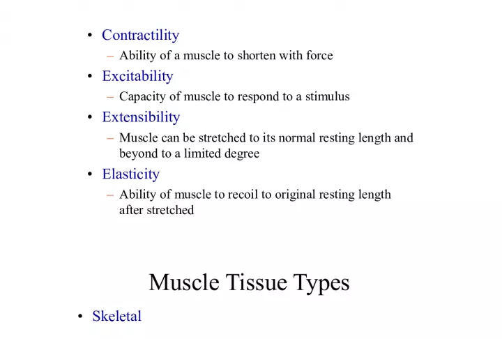
Slide2Properties of Muscle• Contractility – Ability of a muscle to shorten with force • Excitability – Capacity of muscle to respond to a stimulus • Extensibility – Muscle can be stretched to its normal resting length and beyond to a limited degree • Elasticity – Ability of muscle to recoil to original resting length after stretched
Slide3Muscle Tissue Types• Skeletal – Attached to bones – Nuclei multiple and peripherally located – Striated, Voluntary and involuntary (reflexes) • Smooth – Walls of hollow organs, blood vessels, eye, glands, skin – Single nucleus centrally located – Not striated, involuntary • Cardiac – Heart – Single nucleus centrally located – Striations, involuntary, intercalated disks
Slide4Morphology of MuscleFour types: skeletal, cardiac, smooth and myoepithelial cells
Slide5Long multinucleated cells thatrespond only to motor-nerve signals, which cause Ca release from sarcoplasmic reticulum and activation of actin-myosin interaction. Shorter mononucleated cells linked to each other by intercalated disks that contain many gap junctions. Capable of independent, spontaneous contraction, with electrical depolarization transmitted from cell to cell through gap junctions. Spindle-shaped mono- nucleated cells. Contraction influenced by hormones and autonomic nerves. Contraction governed through myosin light chain kinase.
Slide6Skeletal muscle• 40% of adult body weight • 50% of child’s body weight • Muscle contains: – 75% water – 20% protein – 5% organic and inorganic compounds • Functions: – Movement – Maintenance of posture
Slide9Structure of Thick FilamentsMyosin - 2 heavy chains, 4 light chains • Heavy chains - 230 kD • Light chains - 2 pairs of different 20 kD chains • The "heads" of heavy chains have ATPase activity and hydrolysis here drives contraction • Light chains are homologous to calmodulin
Slide10 RyR RyR = ryanodine receptor Ca 2+ channel DHPR DHPR = dihydro- pyridine receptor Ca 2+
Slide13Mechanism of muscle contraction
Slide14Sliding filament model of muscle contraction"Crossbridges" form between myosin and actin, with myosin pulling actin into "H zone" and shortening distance between Z disks.
Slide15Length-tension curve for skeletal muscleFull overlap between thick and thin filaments Decreasing overlap limits maximum tension No overlap (Muscles are not naturally stretched to this point) Actin poking through M line; myosin bumping into Z disk. Contraction range with normal skeletal movements
Slide18Molecular mechanisms of crossbridge action
Slide19T-tubules are NOT positioned at M lines.
Slide20Dihydropyridine ReceptorIn t-tubules of heart and skeletal muscle • Nifedipine and other DHP-like molecules bind to the "DHP receptor" in t-tubules • In heart, DHP receptor is a voltage-gated Ca 2+ channel • In skeletal muscle, DHP receptor is apparently a voltage-sensing protein and probably undergoes voltage-dependent conformational changes
Slide21Ryanodine ReceptorThe "foot structure" in terminal cisternae of SR • Foot structure is a Ca 2+ channel of unusual design • Conformation change or Ca 2+ -channel activity of DHP receptor apparently gates the ryanodine receptor, opening and closing Ca 2+ channels
Slide22Ca2+ Controls Contraction • Release of Ca 2+ from the SR triggers contraction • Reuptake of Ca 2+ into SR relaxes muscle • So how is calcium released in response to nerve impulses? • Answer has come from studies of antagonist molecules that block Ca 2+ channel activity
Slide23This gap isactually only ~10 nm. Ca 2+ -ATPase
Slide25Function of NeuromuscularJunction
Slide26acetate + cholineNa + - - + + end-plate potential (EPP) ~ -15 mV ~ -15mV + + + + + – – – – – ~ +40mV
Slide28Summation of skeletal muscle tension; tetanus
Slide29Contractile forcecan also be regulated through activation of more, or fewer, motor units.
Slide31Muscle contraction• Types – Isometric or static • Constant muscle length • Increased tension – Isotonic • Constant muscle tension • Constant movement » –
Slide33time is required for maximal twitch force to develop, because someshortening of sarcomeres must occur to stretch elastic elements of muscle before force can be transmitted through tendons. By the time this maximal force is developed, [Ca 2+ ] and number of active crossbridges have greatly decreased, so an individual twitch reaches much less than the maximum force the muscle can develop.
Slide34 * Mitochondria generate ~32 ATP from one glucose (slow, but efficient). * Glycolysis generates 2 ATP from one glucose (fast, but inefficient; lactate accumulates). * Creatine kinase reaction: (fastest) ADP + creatine-P ATP + creatine Generate ATP
Slide35Fatigue: * Central — involving central nervous system may involve such factors as dehydration, osmolarity, low blood sugar, and may precede physiological fatigue of actual muscles. * Peripheral — in or near muscles accumulation of lactate and pH, especially in fast-twitch fibers inorganic phosphate — may increasingly inhibit cleavage of ATP in the crossbridge cycle or in the sequestering of Ca 2+ .
Slide36–Incomplete tetanus • Muscle fibers partially relax between contraction • There is time for Ca 2+ to be recycled through the SR between action potentials
Slide37–Complete tetanus • No relaxation between contractions • Action potentials come sp close together that Ca 2+ does not get re- sequestered in the SR
Slide38A skeletal muscle twitch lasts far longer than therefractory period of the stimulating action potential, so many additional stimuli are possible during the twitch, leading to summation of tension and even tetanus.
Slide39in cardiac muscle, the action potential — and thereforethe refractory period — lasts almost as long as the complete muscle contraction, so no tetanus, or even summation, is possible. Sequential contractions are at the same tension, though gradual increases and decreases occur with autonomic nervous system input.
Slide40Copyright © 2008 Pearson Education, Inc., publishing as Benjamin Cummings. Cardiac Muscle Figure 12.37
Slide41Treppe• Graded response • Occurs in muscle rested for prolonged period • Each subsequent contraction is stronger than previous until all equal after few stimuli
Slide43Slow and Fast Fibers:
Slide44Slow and Fast Fibers: